Fluorine in PDB 7m2o: Crystal Structure of Human Glycolate Oxidase with Inhibitor Compound 15
Enzymatic activity of Crystal Structure of Human Glycolate Oxidase with Inhibitor Compound 15
All present enzymatic activity of Crystal Structure of Human Glycolate Oxidase with Inhibitor Compound 15:
1.1.3.15;
1.1.3.15;
Protein crystallography data
The structure of Crystal Structure of Human Glycolate Oxidase with Inhibitor Compound 15, PDB code: 7m2o
was solved by
R.Gumpena,
J.Ding,
D.A.Powell,
W.T.Lowther,
with X-Ray Crystallography technique. A brief refinement statistics is given in the table below:
| Resolution Low / High (Å) | 39.77 / 2.07 |
| Space group | I 4 |
| Cell size a, b, c (Å), α, β, γ (°) | 96.904, 96.904, 79.536, 90, 90, 90 |
| R / Rfree (%) | 12.6 / 16.6 |
Fluorine Binding Sites:
The binding sites of Fluorine atom in the Crystal Structure of Human Glycolate Oxidase with Inhibitor Compound 15
(pdb code 7m2o). This binding sites where shown within
5.0 Angstroms radius around Fluorine atom.
In total 2 binding sites of Fluorine where determined in the Crystal Structure of Human Glycolate Oxidase with Inhibitor Compound 15, PDB code: 7m2o:
Jump to Fluorine binding site number: 1; 2;
In total 2 binding sites of Fluorine where determined in the Crystal Structure of Human Glycolate Oxidase with Inhibitor Compound 15, PDB code: 7m2o:
Jump to Fluorine binding site number: 1; 2;
Fluorine binding site 1 out of 2 in 7m2o
Go back to
Fluorine binding site 1 out
of 2 in the Crystal Structure of Human Glycolate Oxidase with Inhibitor Compound 15
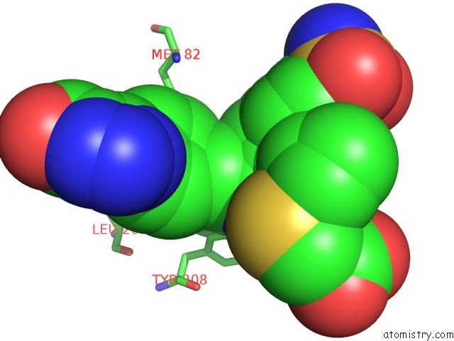
Mono view
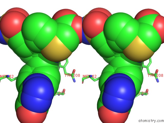
Stereo pair view
Mono view
Stereo pair view
A full contact list of Fluorine with other atoms in the F binding
site number 1 of Crystal Structure of Human Glycolate Oxidase with Inhibitor Compound 15 within 5.0Å range:
|
Fluorine binding site 2 out of 2 in 7m2o
Go back to
Fluorine binding site 2 out
of 2 in the Crystal Structure of Human Glycolate Oxidase with Inhibitor Compound 15
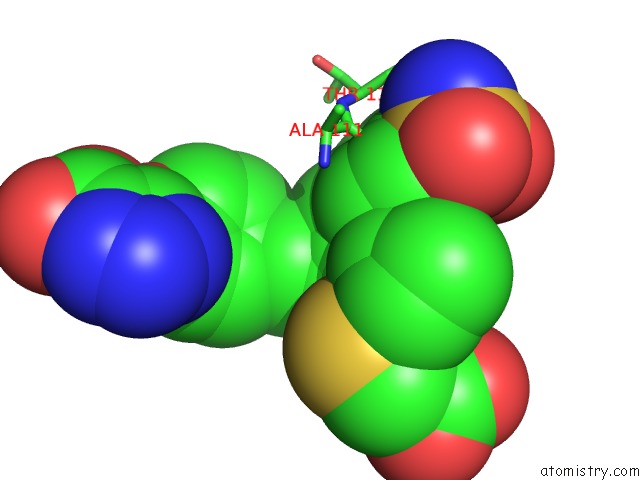
Mono view
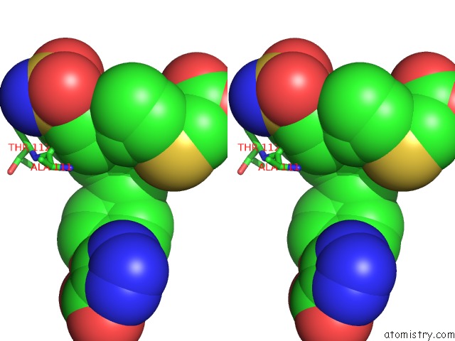
Stereo pair view
Mono view
Stereo pair view
A full contact list of Fluorine with other atoms in the F binding
site number 2 of Crystal Structure of Human Glycolate Oxidase with Inhibitor Compound 15 within 5.0Å range:
|
Reference:
J.Ding,
R.Gumpena,
M.O.Boily,
A.Caron,
O.Chong,
J.H.Cox,
V.Dumais,
S.Gaudreault,
A.H.Graff,
A.King,
J.Knight,
R.Oballa,
J.Surendradoss,
T.Tang,
J.Wu,
W.T.Lowther,
D.A.Powell.
Dual Glycolate Oxidase/Lactate Dehydrogenase A Inhibitors For Primary Hyperoxaluria Acs Medicinal Chemistry V. 12 1116 2021LETTERS.
DOI: 10.1021/ACSMEDCHEMLETT.1C00196
Page generated: Fri Aug 2 09:16:46 2024
DOI: 10.1021/ACSMEDCHEMLETT.1C00196
Last articles
Zn in 9MJ5Zn in 9HNW
Zn in 9G0L
Zn in 9FNE
Zn in 9DZN
Zn in 9E0I
Zn in 9D32
Zn in 9DAK
Zn in 8ZXC
Zn in 8ZUF