Fluorine in PDB 7msk: Thus Glycosin S-Glycosyltransferase
Protein crystallography data
The structure of Thus Glycosin S-Glycosyltransferase, PDB code: 7msk
was solved by
N.Garg,
S.K.Nair,
with X-Ray Crystallography technique. A brief refinement statistics is given in the table below:
| Resolution Low / High (Å) | 25.00 / 2.06 |
| Space group | P 1 21 1 |
| Cell size a, b, c (Å), α, β, γ (°) | 49.833, 112.654, 90.31, 90, 90.11, 90 |
| R / Rfree (%) | 20.1 / 23.9 |
Other elements in 7msk:
The structure of Thus Glycosin S-Glycosyltransferase also contains other interesting chemical elements:
| Magnesium | (Mg) | 2 atoms |
Fluorine Binding Sites:
The binding sites of Fluorine atom in the Thus Glycosin S-Glycosyltransferase
(pdb code 7msk). This binding sites where shown within
5.0 Angstroms radius around Fluorine atom.
In total 2 binding sites of Fluorine where determined in the Thus Glycosin S-Glycosyltransferase, PDB code: 7msk:
Jump to Fluorine binding site number: 1; 2;
In total 2 binding sites of Fluorine where determined in the Thus Glycosin S-Glycosyltransferase, PDB code: 7msk:
Jump to Fluorine binding site number: 1; 2;
Fluorine binding site 1 out of 2 in 7msk
Go back to
Fluorine binding site 1 out
of 2 in the Thus Glycosin S-Glycosyltransferase
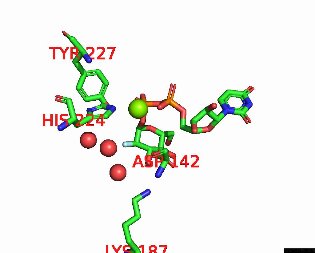
Mono view
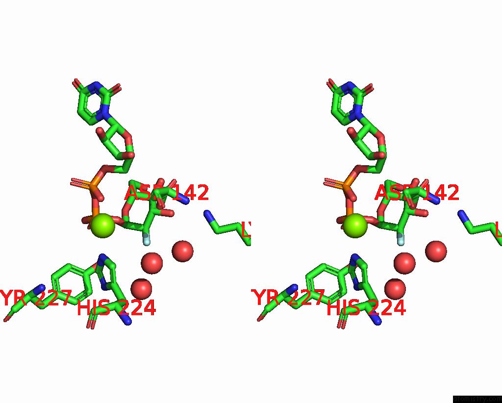
Stereo pair view
Mono view
Stereo pair view
A full contact list of Fluorine with other atoms in the F binding
site number 1 of Thus Glycosin S-Glycosyltransferase within 5.0Å range:
|
Fluorine binding site 2 out of 2 in 7msk
Go back to
Fluorine binding site 2 out
of 2 in the Thus Glycosin S-Glycosyltransferase
Mono view

Stereo pair view
Mono view
Stereo pair view
A full contact list of Fluorine with other atoms in the F binding
site number 2 of Thus Glycosin S-Glycosyltransferase within 5.0Å range:
|
Reference:
D.Fujinami,
C.V.Garcia De Gonzalo,
S.Biswas,
Y.Hao,
H.Wang,
N.Garg,
T.Lukk,
S.K.Nair,
W.A.Van Der Donk.
Structural and Mechanistic Investigations of Protein S-Glycosyltransferases. Cell Chem Biol V. 28 1740 2021.
ISSN: ESSN 2451-9456
PubMed: 34283964
DOI: 10.1016/J.CHEMBIOL.2021.06.009
Page generated: Fri Aug 2 09:47:30 2024
ISSN: ESSN 2451-9456
PubMed: 34283964
DOI: 10.1016/J.CHEMBIOL.2021.06.009
Last articles
Zn in 9MJ5Zn in 9HNW
Zn in 9G0L
Zn in 9FNE
Zn in 9DZN
Zn in 9E0I
Zn in 9D32
Zn in 9DAK
Zn in 8ZXC
Zn in 8ZUF