Fluorine in PDB 7pjc: The Structure of Candida Albicans Phosphoglucomutase with Isothiazolone Modification on CYS359
Enzymatic activity of The Structure of Candida Albicans Phosphoglucomutase with Isothiazolone Modification on CYS359
All present enzymatic activity of The Structure of Candida Albicans Phosphoglucomutase with Isothiazolone Modification on CYS359:
5.4.2.2;
5.4.2.2;
Protein crystallography data
The structure of The Structure of Candida Albicans Phosphoglucomutase with Isothiazolone Modification on CYS359, PDB code: 7pjc
was solved by
K.Yan,
D.M.F.Van Aalten,
with X-Ray Crystallography technique. A brief refinement statistics is given in the table below:
| Resolution Low / High (Å) | 67.97 / 2.11 |
| Space group | P 1 21 1 |
| Cell size a, b, c (Å), α, β, γ (°) | 66.744, 86.444, 110.154, 90, 92.74, 90 |
| R / Rfree (%) | 18.9 / 25.5 |
Other elements in 7pjc:
The structure of The Structure of Candida Albicans Phosphoglucomutase with Isothiazolone Modification on CYS359 also contains other interesting chemical elements:
| Chlorine | (Cl) | 2 atoms |
Fluorine Binding Sites:
The binding sites of Fluorine atom in the The Structure of Candida Albicans Phosphoglucomutase with Isothiazolone Modification on CYS359
(pdb code 7pjc). This binding sites where shown within
5.0 Angstroms radius around Fluorine atom.
In total 2 binding sites of Fluorine where determined in the The Structure of Candida Albicans Phosphoglucomutase with Isothiazolone Modification on CYS359, PDB code: 7pjc:
Jump to Fluorine binding site number: 1; 2;
In total 2 binding sites of Fluorine where determined in the The Structure of Candida Albicans Phosphoglucomutase with Isothiazolone Modification on CYS359, PDB code: 7pjc:
Jump to Fluorine binding site number: 1; 2;
Fluorine binding site 1 out of 2 in 7pjc
Go back to
Fluorine binding site 1 out
of 2 in the The Structure of Candida Albicans Phosphoglucomutase with Isothiazolone Modification on CYS359

Mono view
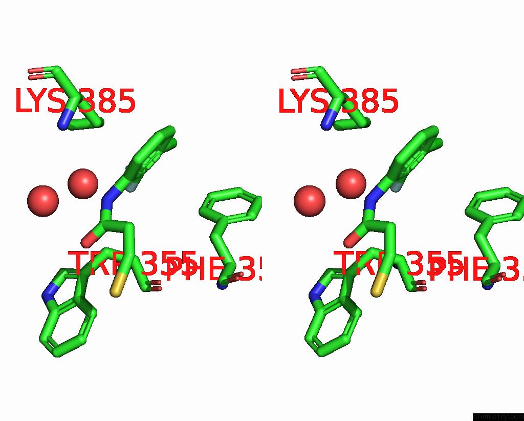
Stereo pair view
Mono view
Stereo pair view
A full contact list of Fluorine with other atoms in the F binding
site number 1 of The Structure of Candida Albicans Phosphoglucomutase with Isothiazolone Modification on CYS359 within 5.0Å range:
|
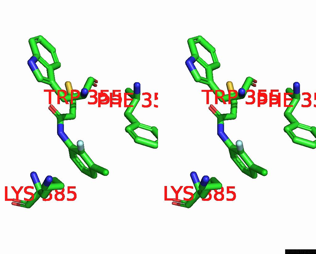
Fluorine binding site 2 out of 2 in 7pjc
Go back to
Fluorine binding site 2 out
of 2 in the The Structure of Candida Albicans Phosphoglucomutase with Isothiazolone Modification on CYS359

Mono view
Stereo pair view
Mono view
Stereo pair view
A full contact list of Fluorine with other atoms in the F binding
site number 2 of The Structure of Candida Albicans Phosphoglucomutase with Isothiazolone Modification on CYS359 within 5.0Å range:
|
Reference:
K.Yan,
M.Stanley,
B.Kowalski,
O.G.Raimi,
A.T.Ferenbach,
P.Wei,
H.Yuan,
W.Fang,
D.M.F.Van Aalten.
Targeting An Essential Step in the Biosynthetic Pathway of Uridine Diphosphate Glucose in Aspergillus Fumigatus To Be Published.
Page generated: Fri Aug 2 11:00:37 2024
Last articles
Zn in 9MJ5Zn in 9HNW
Zn in 9G0L
Zn in 9FNE
Zn in 9DZN
Zn in 9E0I
Zn in 9D32
Zn in 9DAK
Zn in 8ZXC
Zn in 8ZUF