Fluorine in PDB 7rw5: Crystal Structure of Human Methionine Adenosyltransferase 2A (MAT2A) in Complex with Sam and Allosteric Inhibitor Compound 1
Enzymatic activity of Crystal Structure of Human Methionine Adenosyltransferase 2A (MAT2A) in Complex with Sam and Allosteric Inhibitor Compound 1
All present enzymatic activity of Crystal Structure of Human Methionine Adenosyltransferase 2A (MAT2A) in Complex with Sam and Allosteric Inhibitor Compound 1:
2.5.1.6;
2.5.1.6;
Protein crystallography data
The structure of Crystal Structure of Human Methionine Adenosyltransferase 2A (MAT2A) in Complex with Sam and Allosteric Inhibitor Compound 1, PDB code: 7rw5
was solved by
L.Jin,
A.K.Padyana,
with X-Ray Crystallography technique. A brief refinement statistics is given in the table below:
| Resolution Low / High (Å) | 37.92 / 2.48 |
| Space group | P 1 21 1 |
| Cell size a, b, c (Å), α, β, γ (°) | 68.237, 103.99, 113.972, 90, 93.47, 90 |
| R / Rfree (%) | 17.5 / 22.9 |
Fluorine Binding Sites:
The binding sites of Fluorine atom in the Crystal Structure of Human Methionine Adenosyltransferase 2A (MAT2A) in Complex with Sam and Allosteric Inhibitor Compound 1
(pdb code 7rw5). This binding sites where shown within
5.0 Angstroms radius around Fluorine atom.
In total only one binding site of Fluorine was determined in the Crystal Structure of Human Methionine Adenosyltransferase 2A (MAT2A) in Complex with Sam and Allosteric Inhibitor Compound 1, PDB code: 7rw5:
In total only one binding site of Fluorine was determined in the Crystal Structure of Human Methionine Adenosyltransferase 2A (MAT2A) in Complex with Sam and Allosteric Inhibitor Compound 1, PDB code: 7rw5:
Fluorine binding site 1 out of 1 in 7rw5
Go back to
Fluorine binding site 1 out
of 1 in the Crystal Structure of Human Methionine Adenosyltransferase 2A (MAT2A) in Complex with Sam and Allosteric Inhibitor Compound 1
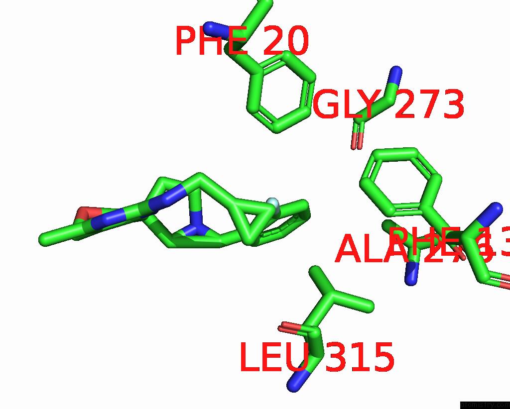
Mono view
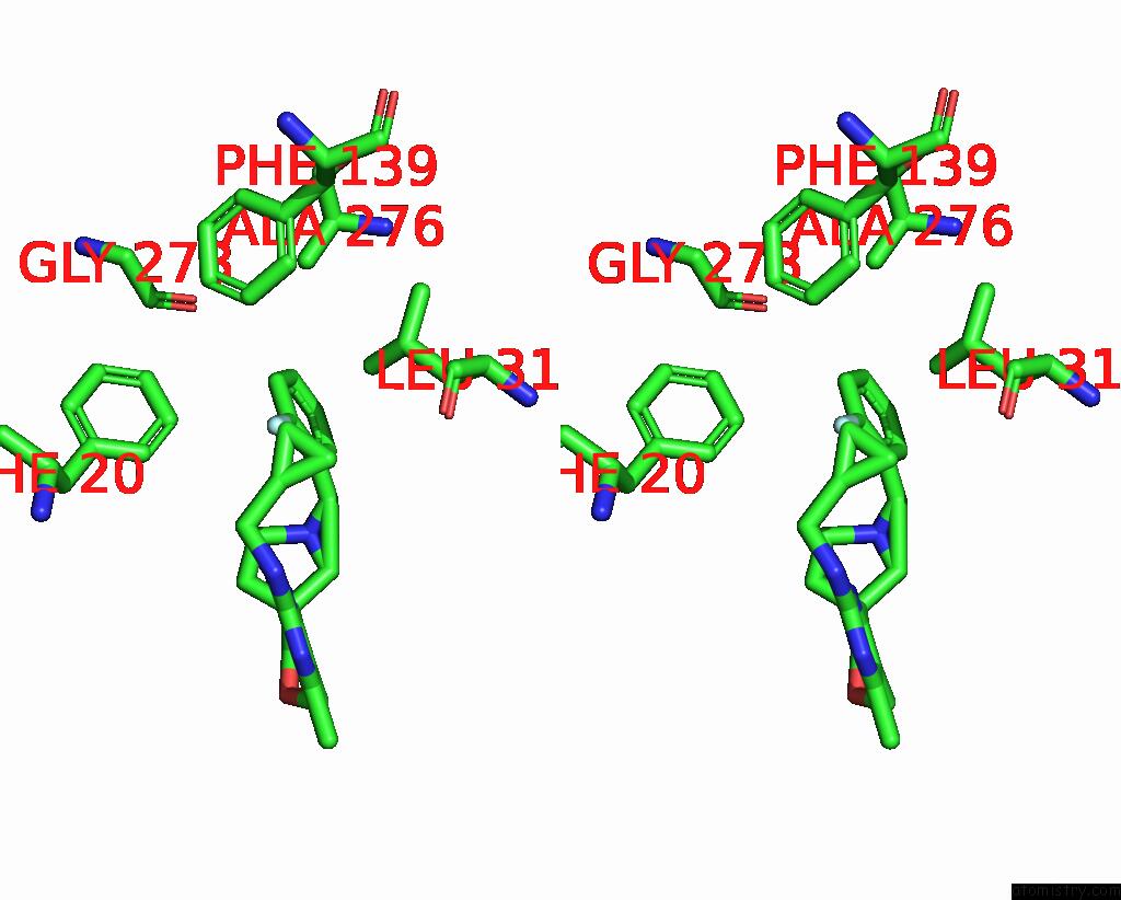
Stereo pair view
Mono view
Stereo pair view
A full contact list of Fluorine with other atoms in the F binding
site number 1 of Crystal Structure of Human Methionine Adenosyltransferase 2A (MAT2A) in Complex with Sam and Allosteric Inhibitor Compound 1 within 5.0Å range:
|
Reference:
M.Li,
Z.Konteatis,
N.Nagaraja,
Y.Chen,
S.Zhou,
G.Ma,
S.Gross,
K.Marjon,
M.L.Hyer,
E.Mandley,
M.Lein,
A.K.Padyana,
L.Jin,
S.Tong,
R.Peters,
J.Murtie,
J.Travins,
M.Medeiros,
P.Liu,
V.Frank,
E.T.Judd,
S.A.Biller,
K.M.Marks,
Z.Sui,
S.K.Reznik.
Leveraging Structure-Based Drug Design to Identify Next-Generation MAT2A Inhibitors, Including Brain-Penetrant and Peripherally Efficacious Leads. J.Med.Chem. V. 65 4600 2022.
ISSN: ISSN 0022-2623
PubMed: 35293760
DOI: 10.1021/ACS.JMEDCHEM.1C01595
Page generated: Fri Aug 2 12:36:07 2024
ISSN: ISSN 0022-2623
PubMed: 35293760
DOI: 10.1021/ACS.JMEDCHEM.1C01595
Last articles
Zn in 9MJ5Zn in 9HNW
Zn in 9G0L
Zn in 9FNE
Zn in 9DZN
Zn in 9E0I
Zn in 9D32
Zn in 9DAK
Zn in 8ZXC
Zn in 8ZUF