Fluorine in PDB 7sed: Crystal Structure of Human Fibrillarin in Complex with Compound 2A

Protein crystallography data

The structure of Crystal Structure of Human Fibrillarin in Complex with Compound 2A, PDB code: 7sed was solved by Y.Shi, I.M.El-Deeb, V.Masic, L.Hartley-Tassell, A.Maggioni, M.Vonitzstein, T.Ve, with X-Ray Crystallography technique. A brief refinement statistics is given in the table below:

Resolution Low / High (Å) 38.63 / 1.90
Space group C 2 2 21
Cell size a, b, c (Å), α, β, γ (°) 69.291, 139.581, 66.249, 90, 90, 90
R / Rfree (%) 18.9 / 22.5

Fluorine Binding Sites:

The binding sites of Fluorine atom in the Crystal Structure of Human Fibrillarin in Complex with Compound 2A (pdb code 7sed). This binding sites where shown within 5.0 Angstroms radius around Fluorine atom.
In total 3 binding sites of Fluorine where determined in the Crystal Structure of Human Fibrillarin in Complex with Compound 2A, PDB code: 7sed:
Jump to Fluorine binding site number: 1; 2; 3;

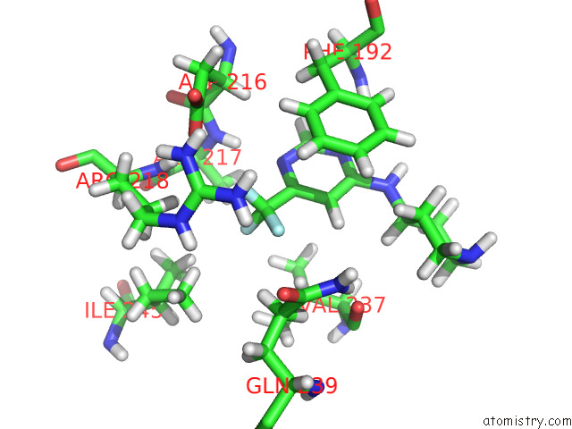
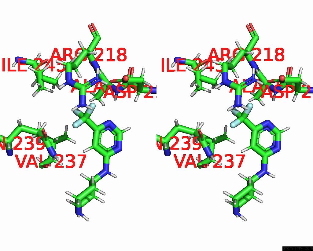
Fluorine binding site 1 out of 3 in 7sed

Go back to Fluorine Binding Sites List in 7sed
Fluorine binding site 1 out of 3 in the Crystal Structure of Human Fibrillarin in Complex with Compound 2A


Mono view


Stereo pair view

A full contact list of Fluorine with other atoms in the F binding site number 1 of Crystal Structure of Human Fibrillarin in Complex with Compound 2A within 5.0Å range:
probe atom residue distance (Å) B Occ
A:F401

b:42.3
occ:1.00
F A:8VD401 0.0 42.3 1.0
C A:8VD401 1.3 39.5 1.0
F1 A:8VD401 2.1 38.2 1.0
F2 A:8VD401 2.1 44.0 1.0
C1 A:8VD401 2.3 34.6 1.0
OD1 A:ASP216 2.6 34.8 1.0
HG3 A:ARG218 2.6 47.5 1.0
N3 A:8VD401 2.7 28.8 1.0
H A:ALA217 3.0 31.6 1.0
H A:ARG218 3.1 33.5 1.0
HB3 A:ALA217 3.5 32.6 1.0
C2 A:8VD401 3.5 34.8 1.0
CG A:ASP216 3.6 39.9 1.0
N A:ALA217 3.6 26.3 1.0
HD2 A:PHE192 3.6 61.5 1.0
CZ A:ARG218 3.6 56.4 1.0
CG A:ARG218 3.6 39.5 1.0
HE2 A:PHE192 3.6 57.5 1.0
HD13 A:ILE245 3.7 44.8 1.0
HA A:ASP216 3.7 37.9 1.0
N A:ARG218 3.7 27.9 1.0
NH2 A:ARG218 3.8 65.2 1.0
NE A:ARG218 3.8 50.0 1.0
H A:8VD401 3.9 41.8 1.0
CD2 A:PHE192 3.9 51.2 1.0
CE2 A:PHE192 3.9 47.8 1.0
NH1 A:ARG218 3.9 59.9 1.0
HH21 A:ARG218 4.0 78.4 1.0
C9 A:8VD401 4.0 27.0 1.0
HG2 A:ARG218 4.0 47.5 1.0
HE A:ARG218 4.0 60.1 1.0
HH22 A:ARG218 4.0 78.4 1.0
OD2 A:ASP216 4.2 45.6 1.0
HH12 A:ARG218 4.2 71.9 1.0
HH11 A:ARG218 4.2 71.9 1.0
HG3 A:GLN239 4.2 51.6 1.0
C A:ASP216 4.2 26.6 1.0
CA A:ALA217 4.2 24.9 1.0
CB A:ALA217 4.3 27.1 1.0
CA A:ASP216 4.3 31.5 1.0
CD A:ARG218 4.3 49.8 1.0
C A:ALA217 4.4 28.7 1.0
HB2 A:ARG218 4.4 38.0 1.0
HG21 A:ILE245 4.4 36.0 1.0
CB A:ARG218 4.5 31.6 1.0
CB A:ASP216 4.5 36.0 1.0
HB A:VAL237 4.6 48.7 1.0
H12 A:8VD401 4.6 32.5 1.0
C3 A:8VD401 4.6 33.1 1.0
HB2 A:ALA217 4.6 32.6 1.0
CD1 A:ILE245 4.6 37.2 1.0
CA A:ARG218 4.7 29.2 1.0
HD11 A:ILE245 4.8 44.8 1.0
N2 A:8VD401 4.8 30.3 1.0
HD2 A:ARG218 4.8 59.9 1.0
HB3 A:PHE192 4.8 42.8 1.0
HG11 A:VAL237 4.8 43.4 1.0
CG A:PHE192 4.9 42.6 1.0
CZ A:PHE192 4.9 49.9 1.0
HA A:ARG218 5.0 35.1 1.0

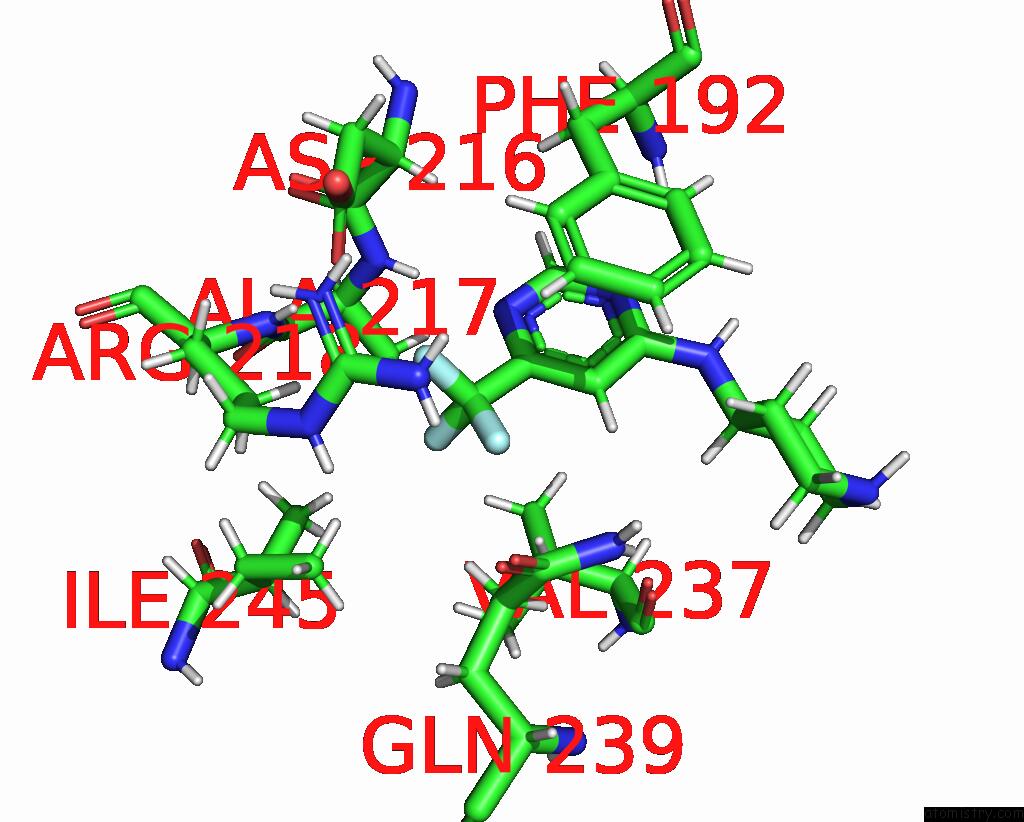
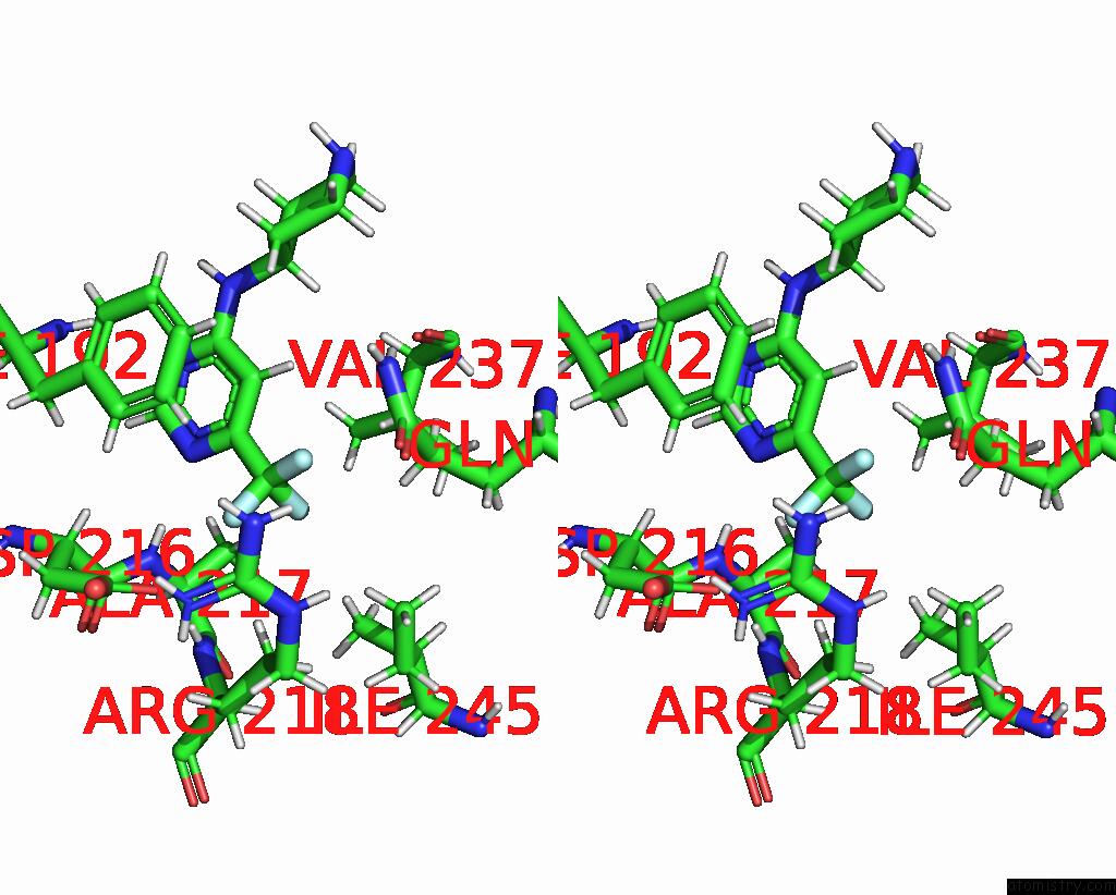
Fluorine binding site 2 out of 3 in 7sed

Go back to Fluorine Binding Sites List in 7sed
Fluorine binding site 2 out of 3 in the Crystal Structure of Human Fibrillarin in Complex with Compound 2A


Mono view


Stereo pair view

A full contact list of Fluorine with other atoms in the F binding site number 2 of Crystal Structure of Human Fibrillarin in Complex with Compound 2A within 5.0Å range:
probe atom residue distance (Å) B Occ
A:F401

b:38.2
occ:1.00
F1 A:8VD401 0.0 38.2 1.0
C A:8VD401 1.3 39.5 1.0
F A:8VD401 2.1 42.3 1.0
F2 A:8VD401 2.1 44.0 1.0
C1 A:8VD401 2.3 34.6 1.0
HB3 A:ALA217 2.5 32.6 1.0
HD13 A:ILE245 2.7 44.8 1.0
HG11 A:VAL237 2.9 43.4 1.0
HG21 A:ILE245 2.9 36.0 1.0
HB A:VAL237 2.9 48.7 1.0
N3 A:8VD401 3.0 28.8 1.0
C2 A:8VD401 3.3 34.8 1.0
CB A:ALA217 3.5 27.1 1.0
CG1 A:VAL237 3.5 36.0 1.0
HG3 A:ARG218 3.5 47.5 1.0
H A:ALA217 3.5 31.6 1.0
HG12 A:VAL237 3.5 43.4 1.0
H A:8VD401 3.5 41.8 1.0
CB A:VAL237 3.6 40.5 1.0
HG3 A:GLN239 3.6 51.6 1.0
HG21 A:VAL237 3.7 45.1 1.0
CD1 A:ILE245 3.7 37.2 1.0
H A:ARG218 3.8 33.5 1.0
CG2 A:ILE245 3.9 30.0 1.0
HB2 A:ALA217 3.9 32.6 1.0
HD12 A:ILE245 3.9 44.8 1.0
HB1 A:ALA217 4.0 32.6 1.0
N A:ALA217 4.1 26.3 1.0
CA A:ALA217 4.1 24.9 1.0
N A:ARG218 4.1 27.9 1.0
HG2 A:ARG218 4.1 47.5 1.0
CG2 A:VAL237 4.2 37.5 1.0
HD11 A:ILE245 4.2 44.8 1.0
HG22 A:ILE245 4.2 36.0 1.0
C9 A:8VD401 4.2 27.0 1.0
CG A:ARG218 4.3 39.5 1.0
C A:ALA217 4.3 28.7 1.0
HG23 A:ILE245 4.3 36.0 1.0
OD1 A:ASP216 4.3 34.8 1.0
HG13 A:VAL237 4.4 43.4 1.0
HG2 A:GLN239 4.4 51.6 1.0
CG A:GLN239 4.4 43.0 1.0
HG12 A:ILE245 4.5 37.0 1.0
C3 A:8VD401 4.5 33.1 1.0
CG1 A:ILE245 4.5 30.8 1.0
HG23 A:VAL237 4.6 45.1 1.0
CB A:ILE245 4.7 27.1 1.0
HB A:ILE245 4.7 32.6 1.0
HE A:ARG218 4.8 60.1 1.0
NE A:ARG218 4.9 50.0 1.0
N2 A:8VD401 4.9 30.3 1.0
CA A:VAL237 4.9 35.8 1.0
HG22 A:VAL237 4.9 45.1 1.0
HA A:VAL237 4.9 43.1 1.0
H12 A:8VD401 4.9 32.5 1.0
HA A:ARG218 5.0 35.1 1.0

Fluorine binding site 3 out of 3 in 7sed

Go back to Fluorine Binding Sites List in 7sed
Fluorine binding site 3 out of 3 in the Crystal Structure of Human Fibrillarin in Complex with Compound 2A


Mono view


Stereo pair view

A full contact list of Fluorine with other atoms in the F binding site number 3 of Crystal Structure of Human Fibrillarin in Complex with Compound 2A within 5.0Å range:
probe atom residue distance (Å) B Occ
A:F401

b:44.0
occ:1.00
F2 A:8VD401 0.0 44.0 1.0
C A:8VD401 1.3 39.5 1.0
F A:8VD401 2.1 42.3 1.0
F1 A:8VD401 2.1 38.2 1.0
C1 A:8VD401 2.4 34.6 1.0
H A:8VD401 2.5 41.8 1.0
HG3 A:GLN239 2.5 51.6 1.0
C2 A:8VD401 2.8 34.8 1.0
HB A:VAL237 3.1 48.7 1.0
CG A:GLN239 3.2 43.0 1.0
HG2 A:GLN239 3.3 51.6 1.0
CD A:GLN239 3.4 50.5 1.0
HE21 A:GLN239 3.5 67.0 1.0
N3 A:8VD401 3.5 28.8 1.0
NE2 A:GLN239 3.5 55.7 1.0
HH21 A:ARG218 3.6 78.4 1.0
HE2 A:PHE192 3.6 57.5 1.0
HD13 A:ILE245 3.7 44.8 1.0
NH2 A:ARG218 3.8 65.2 1.0
CE2 A:PHE192 3.9 47.8 1.0
HG3 A:ARG218 4.0 47.5 1.0
CB A:VAL237 4.0 40.5 1.0
OE1 A:GLN239 4.0 42.3 1.0
HE A:ARG218 4.0 60.1 1.0
HE22 A:GLN239 4.0 67.0 1.0
C3 A:8VD401 4.1 33.1 1.0
HH22 A:ARG218 4.2 78.4 1.0
O A:VAL237 4.2 39.4 1.0
CZ A:ARG218 4.2 56.4 1.0
NE A:ARG218 4.3 50.0 1.0
HG11 A:VAL237 4.3 43.4 1.0
CZ A:PHE192 4.4 49.9 1.0
HG12 A:VAL237 4.4 43.4 1.0
HZ A:PHE192 4.4 59.9 1.0
CD1 A:ILE245 4.4 37.2 1.0
HB3 A:ALA217 4.5 32.6 1.0
CG1 A:VAL237 4.5 36.0 1.0
CD2 A:PHE192 4.5 51.2 1.0
H3 A:8VD401 4.5 53.9 1.0
HD11 A:ILE245 4.5 44.8 1.0
HG21 A:VAL237 4.5 45.1 1.0
HD12 A:ILE245 4.5 44.8 1.0
C9 A:8VD401 4.6 27.0 1.0
CB A:GLN239 4.6 44.0 1.0
H2 A:8VD401 4.6 45.4 1.0
HD2 A:PHE192 4.6 61.5 1.0
OD1 A:ASP216 4.7 34.8 1.0
H A:ALA217 4.7 31.6 1.0
HB2 A:GLN239 4.7 52.9 1.0
CG2 A:VAL237 4.7 37.5 1.0
HG21 A:ILE245 4.8 36.0 1.0
CG A:ARG218 4.8 39.5 1.0
HG23 A:VAL237 4.8 45.1 1.0
HA A:VAL237 4.8 43.1 1.0
HB3 A:GLN239 4.8 52.9 1.0
CA A:VAL237 4.9 35.8 1.0
N2 A:8VD401 4.9 30.3 1.0
C A:VAL237 4.9 42.5 1.0
HG2 A:ARG218 4.9 47.5 1.0

Reference:

Y.Shi, I.M.El-Deeb, V.Masic, L.Hartley-Tassell, A.Maggioni, M.V.Itzstein, T.Ve. Discovery of Cofactor Competitive Inhibitors Against the Human Methyltransferase Fibrillarin. Pharmaceuticals V. 15 2021.
ISSN: ESSN 1424-8247
PubMed: 35056083
DOI: 10.3390/PH15010026
Page generated: Fri Aug 2 12:53:58 2024

Last articles

Zn in 9MJ5
Zn in 9HNW
Zn in 9G0L
Zn in 9FNE
Zn in 9DZN
Zn in 9E0I
Zn in 9D32
Zn in 9DAK
Zn in 8ZXC
Zn in 8ZUF
© Copyright 2008-2020 by atomistry.com
Home   |    Site Map   |    Copyright   |    Contact us   |    Privacy