Fluorine in PDB 8a19: Structure of A Leucinostatin Derivative Determined By Host Lattice Display : L1E4V1 Construct
Protein crystallography data
The structure of Structure of A Leucinostatin Derivative Determined By Host Lattice Display : L1E4V1 Construct, PDB code: 8a19
was solved by
P.R.E.Mittl,
with X-Ray Crystallography technique. A brief refinement statistics is given in the table below:
| Resolution Low / High (Å) | 34.52 / 2.36 |
| Space group | P 65 |
| Cell size a, b, c (Å), α, β, γ (°) | 192.757, 192.757, 122.834, 90, 90, 120 |
| R / Rfree (%) | 16.3 / 19.3 |
Other elements in 8a19:
The structure of Structure of A Leucinostatin Derivative Determined By Host Lattice Display : L1E4V1 Construct also contains other interesting chemical elements:
| Manganese | (Mn) | 4 atoms |
| Chlorine | (Cl) | 5 atoms |
Fluorine Binding Sites:
The binding sites of Fluorine atom in the Structure of A Leucinostatin Derivative Determined By Host Lattice Display : L1E4V1 Construct
(pdb code 8a19). This binding sites where shown within
5.0 Angstroms radius around Fluorine atom.
In total only one binding site of Fluorine was determined in the Structure of A Leucinostatin Derivative Determined By Host Lattice Display : L1E4V1 Construct, PDB code: 8a19:
In total only one binding site of Fluorine was determined in the Structure of A Leucinostatin Derivative Determined By Host Lattice Display : L1E4V1 Construct, PDB code: 8a19:
Fluorine binding site 1 out of 1 in 8a19
Go back to
Fluorine binding site 1 out
of 1 in the Structure of A Leucinostatin Derivative Determined By Host Lattice Display : L1E4V1 Construct

Mono view
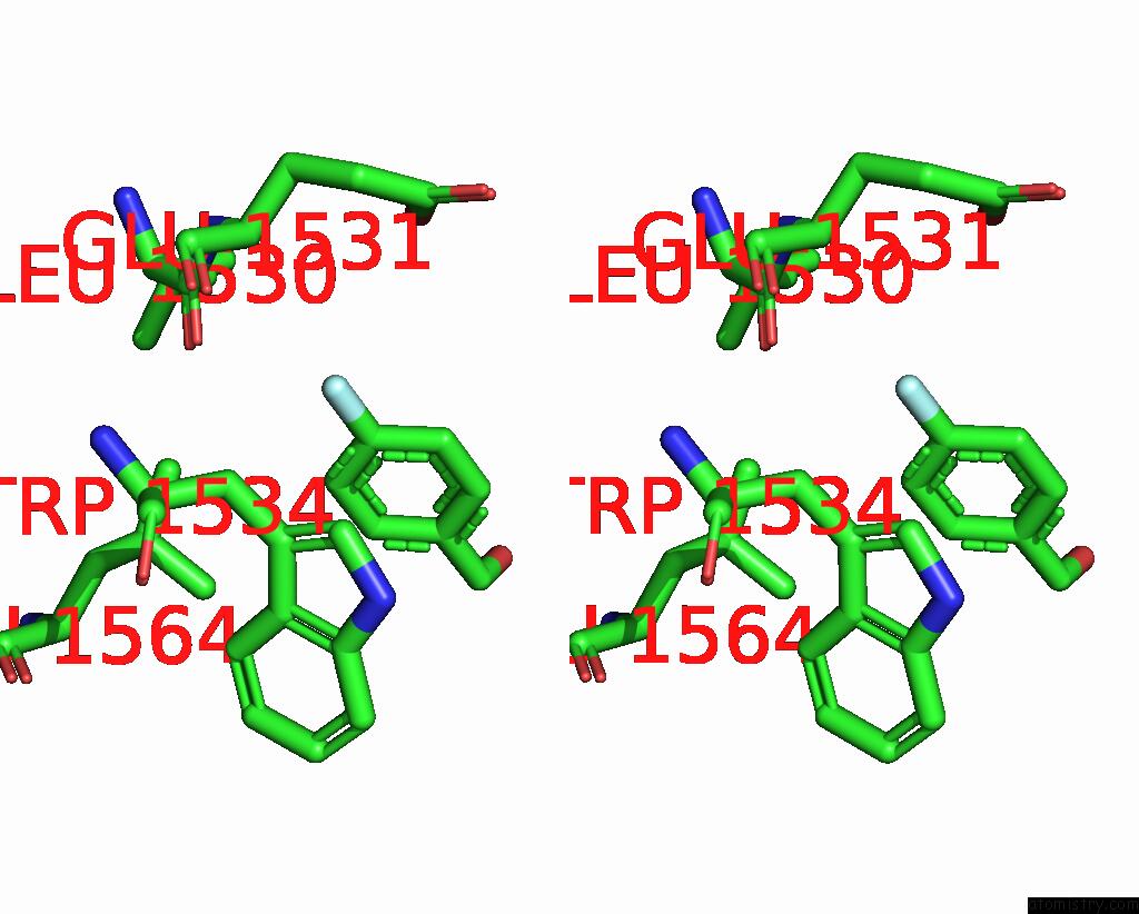
Stereo pair view
Mono view
Stereo pair view
A full contact list of Fluorine with other atoms in the F binding
site number 1 of Structure of A Leucinostatin Derivative Determined By Host Lattice Display : L1E4V1 Construct within 5.0Å range:
|
Reference:
C.Kiss,
F.M.Gall,
B.Dreier,
M.Adams,
R.Riedl,
A.Pluckthun,
P.R.E.Mittl.
Structure of A Hydrophobic Leucinostatin Derivative Determined By Host Lattice Display. Acta Crystallogr D Struct V. 78 1439 2022BIOL.
ISSN: ISSN 2059-7983
PubMed: 36458615
DOI: 10.1107/S2059798322010762
Page generated: Fri Aug 2 16:23:05 2024
ISSN: ISSN 2059-7983
PubMed: 36458615
DOI: 10.1107/S2059798322010762
Last articles
Zn in 9JYWZn in 9IR4
Zn in 9IR3
Zn in 9GMX
Zn in 9GMW
Zn in 9JEJ
Zn in 9ERF
Zn in 9ERE
Zn in 9EGV
Zn in 9EGW