Fluorine in PDB 8c0r: Crystal Structure of Human Carbonic Anhydrase II in Complex with A Coumarin Derivative.
Enzymatic activity of Crystal Structure of Human Carbonic Anhydrase II in Complex with A Coumarin Derivative.
All present enzymatic activity of Crystal Structure of Human Carbonic Anhydrase II in Complex with A Coumarin Derivative.:
4.2.1.1;
4.2.1.1;
Protein crystallography data
The structure of Crystal Structure of Human Carbonic Anhydrase II in Complex with A Coumarin Derivative., PDB code: 8c0r
was solved by
V.Alterio,
G.De Simone,
D.Esposito,
with X-Ray Crystallography technique. A brief refinement statistics is given in the table below:
| Resolution Low / High (Å) | 24.60 / 1.56 |
| Space group | P 1 21 1 |
| Cell size a, b, c (Å), α, β, γ (°) | 42.342, 41.484, 72.053, 90, 104.38, 90 |
| R / Rfree (%) | 17.7 / 20.1 |
Other elements in 8c0r:
The structure of Crystal Structure of Human Carbonic Anhydrase II in Complex with A Coumarin Derivative. also contains other interesting chemical elements:
| Zinc | (Zn) | 1 atom |
Fluorine Binding Sites:
The binding sites of Fluorine atom in the Crystal Structure of Human Carbonic Anhydrase II in Complex with A Coumarin Derivative.
(pdb code 8c0r). This binding sites where shown within
5.0 Angstroms radius around Fluorine atom.
In total 2 binding sites of Fluorine where determined in the Crystal Structure of Human Carbonic Anhydrase II in Complex with A Coumarin Derivative., PDB code: 8c0r:
Jump to Fluorine binding site number: 1; 2;
In total 2 binding sites of Fluorine where determined in the Crystal Structure of Human Carbonic Anhydrase II in Complex with A Coumarin Derivative., PDB code: 8c0r:
Jump to Fluorine binding site number: 1; 2;
Fluorine binding site 1 out of 2 in 8c0r
Go back to
Fluorine binding site 1 out
of 2 in the Crystal Structure of Human Carbonic Anhydrase II in Complex with A Coumarin Derivative.
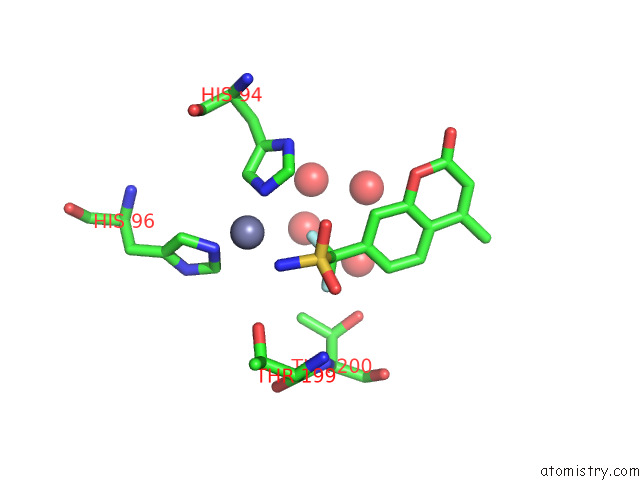
Mono view
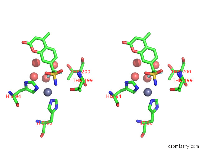
Stereo pair view
Mono view
Stereo pair view
A full contact list of Fluorine with other atoms in the F binding
site number 1 of Crystal Structure of Human Carbonic Anhydrase II in Complex with A Coumarin Derivative. within 5.0Å range:
|
Fluorine binding site 2 out of 2 in 8c0r
Go back to
Fluorine binding site 2 out
of 2 in the Crystal Structure of Human Carbonic Anhydrase II in Complex with A Coumarin Derivative.
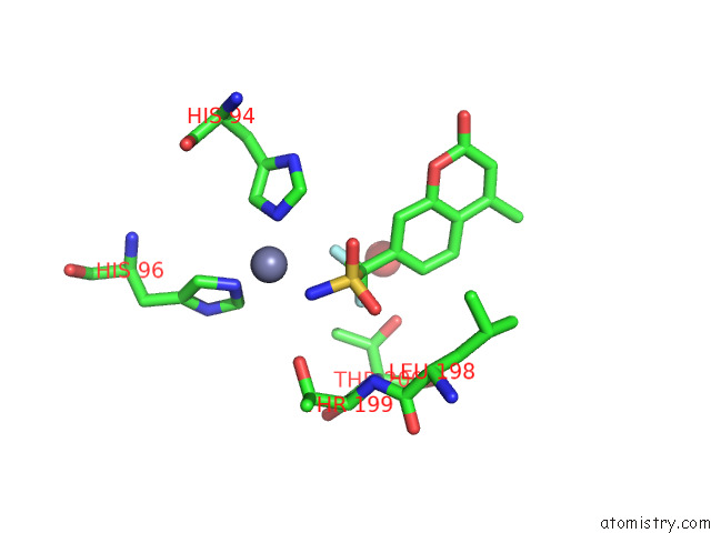
Mono view
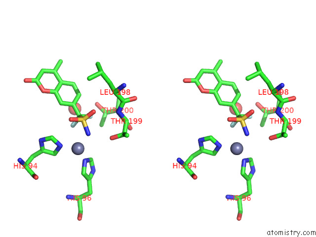
Stereo pair view
Mono view
Stereo pair view
A full contact list of Fluorine with other atoms in the F binding
site number 2 of Crystal Structure of Human Carbonic Anhydrase II in Complex with A Coumarin Derivative. within 5.0Å range:
|
Reference:
E.Langella,
D.Esposito,
S.M.Monti,
C.T.Supuran,
G.De Simone,
V.Alterio.
A Combined in Silico and Structural Study Opens New Perspectives on Aliphatic Sulfonamides, A Still Poorly Investigated Class of Ca Inhibitors. Biology (Basel) V. 12 2023.
ISSN: ESSN 2079-7737
PubMed: 36829558
DOI: 10.3390/BIOLOGY12020281
Page generated: Fri Aug 2 17:01:30 2024
ISSN: ESSN 2079-7737
PubMed: 36829558
DOI: 10.3390/BIOLOGY12020281
Last articles
Zn in 9MJ5Zn in 9HNW
Zn in 9G0L
Zn in 9FNE
Zn in 9DZN
Zn in 9E0I
Zn in 9D32
Zn in 9DAK
Zn in 8ZXC
Zn in 8ZUF