Fluorine in PDB 8f57: Crystal Structure of Acetyltransferase Eis From M. Tuberculosis in Complex with Inhibitor SGT1615
Protein crystallography data
The structure of Crystal Structure of Acetyltransferase Eis From M. Tuberculosis in Complex with Inhibitor SGT1615, PDB code: 8f57
was solved by
A.H.Pang,
A.Punetha,
S.Garneau-Tsodikova,
O.V.Tsodikov,
with X-Ray Crystallography technique. A brief refinement statistics is given in the table below:
| Resolution Low / High (Å) | 34.70 / 2.42 |
| Space group | H 3 2 |
| Cell size a, b, c (Å), α, β, γ (°) | 174.907, 174.907, 123.252, 90, 90, 120 |
| R / Rfree (%) | 17.4 / 20.5 |
Other elements in 8f57:
The structure of Crystal Structure of Acetyltransferase Eis From M. Tuberculosis in Complex with Inhibitor SGT1615 also contains other interesting chemical elements:
| Chlorine | (Cl) | 1 atom |
Fluorine Binding Sites:
The binding sites of Fluorine atom in the Crystal Structure of Acetyltransferase Eis From M. Tuberculosis in Complex with Inhibitor SGT1615
(pdb code 8f57). This binding sites where shown within
5.0 Angstroms radius around Fluorine atom.
In total only one binding site of Fluorine was determined in the Crystal Structure of Acetyltransferase Eis From M. Tuberculosis in Complex with Inhibitor SGT1615, PDB code: 8f57:
In total only one binding site of Fluorine was determined in the Crystal Structure of Acetyltransferase Eis From M. Tuberculosis in Complex with Inhibitor SGT1615, PDB code: 8f57:
Fluorine binding site 1 out of 1 in 8f57
Go back to
Fluorine binding site 1 out
of 1 in the Crystal Structure of Acetyltransferase Eis From M. Tuberculosis in Complex with Inhibitor SGT1615

Mono view
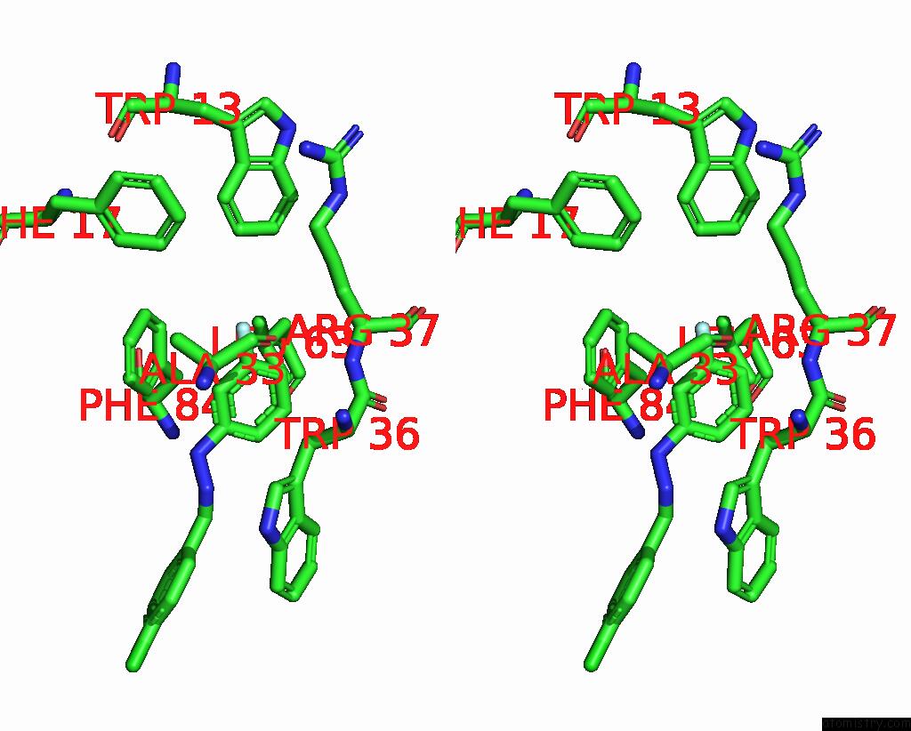
Stereo pair view
Mono view
Stereo pair view
A full contact list of Fluorine with other atoms in the F binding
site number 1 of Crystal Structure of Acetyltransferase Eis From M. Tuberculosis in Complex with Inhibitor SGT1615 within 5.0Å range:
|
Reference:
A.H.Pang,
K.D.Green,
A.Punetha,
N.Thamban Chandrika,
K.C.Howard,
S.Garneau-Tsodikova,
O.V.Tsodikov.
Discovery and Mechanistic Analysis of Structurally Diverse Inhibitors of Acetyltransferase Eis Among Fda-Approved Drugs. Biochemistry 2023.
ISSN: ISSN 0006-2960
PubMed: 36657084
DOI: 10.1021/ACS.BIOCHEM.2C00658
Page generated: Fri Aug 2 18:31:53 2024
ISSN: ISSN 0006-2960
PubMed: 36657084
DOI: 10.1021/ACS.BIOCHEM.2C00658
Last articles
Zn in 9MJ5Zn in 9HNW
Zn in 9G0L
Zn in 9FNE
Zn in 9DZN
Zn in 9E0I
Zn in 9D32
Zn in 9DAK
Zn in 8ZXC
Zn in 8ZUF