Fluorine »
PDB 9f2l-9mel »
9gla »
Fluorine in PDB 9gla: Crystal Structure of A CDK2-Based CDK7 Mimic with Inhibitor SY5609
Enzymatic activity of Crystal Structure of A CDK2-Based CDK7 Mimic with Inhibitor SY5609
All present enzymatic activity of Crystal Structure of A CDK2-Based CDK7 Mimic with Inhibitor SY5609:
2.7.11.22;
2.7.11.22;
Protein crystallography data
The structure of Crystal Structure of A CDK2-Based CDK7 Mimic with Inhibitor SY5609, PDB code: 9gla
was solved by
J.Skerlova,
V.Krejcirikova,
P.Rezacova,
with X-Ray Crystallography technique. A brief refinement statistics is given in the table below:
| Resolution Low / High (Å) | 48.46 / 2.18 |
| Space group | C 2 2 21 |
| Cell size a, b, c (Å), α, β, γ (°) | 71.937, 109.786, 163.554, 90, 90, 90 |
| R / Rfree (%) | 20.1 / 24.9 |
Other elements in 9gla:
The structure of Crystal Structure of A CDK2-Based CDK7 Mimic with Inhibitor SY5609 also contains other interesting chemical elements:
| Sodium | (Na) | 2 atoms |
Fluorine Binding Sites:
The binding sites of Fluorine atom in the Crystal Structure of A CDK2-Based CDK7 Mimic with Inhibitor SY5609
(pdb code 9gla). This binding sites where shown within
5.0 Angstroms radius around Fluorine atom.
In total 3 binding sites of Fluorine where determined in the Crystal Structure of A CDK2-Based CDK7 Mimic with Inhibitor SY5609, PDB code: 9gla:
Jump to Fluorine binding site number: 1; 2; 3;
In total 3 binding sites of Fluorine where determined in the Crystal Structure of A CDK2-Based CDK7 Mimic with Inhibitor SY5609, PDB code: 9gla:
Jump to Fluorine binding site number: 1; 2; 3;
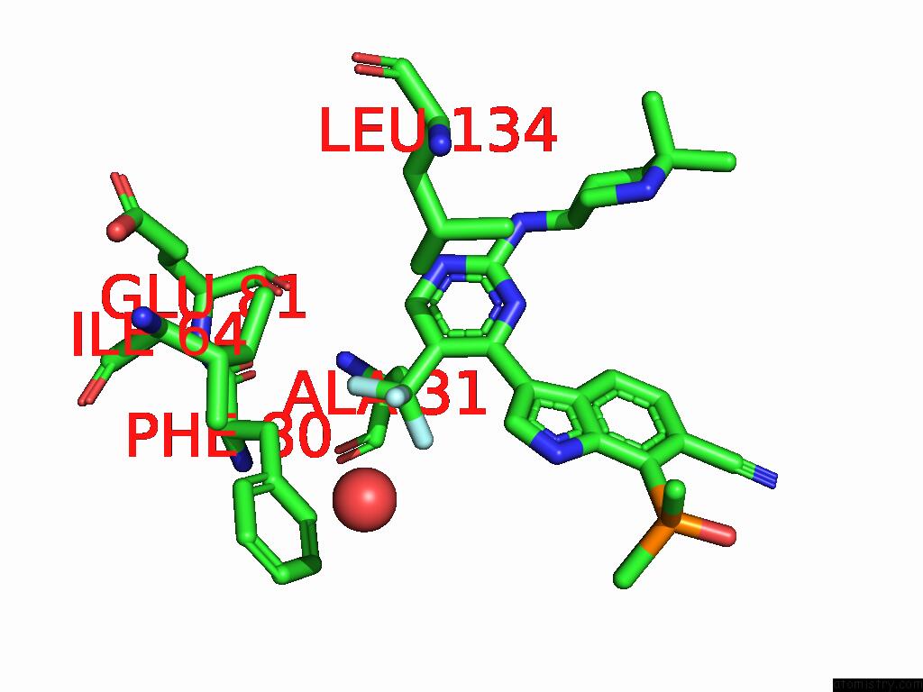
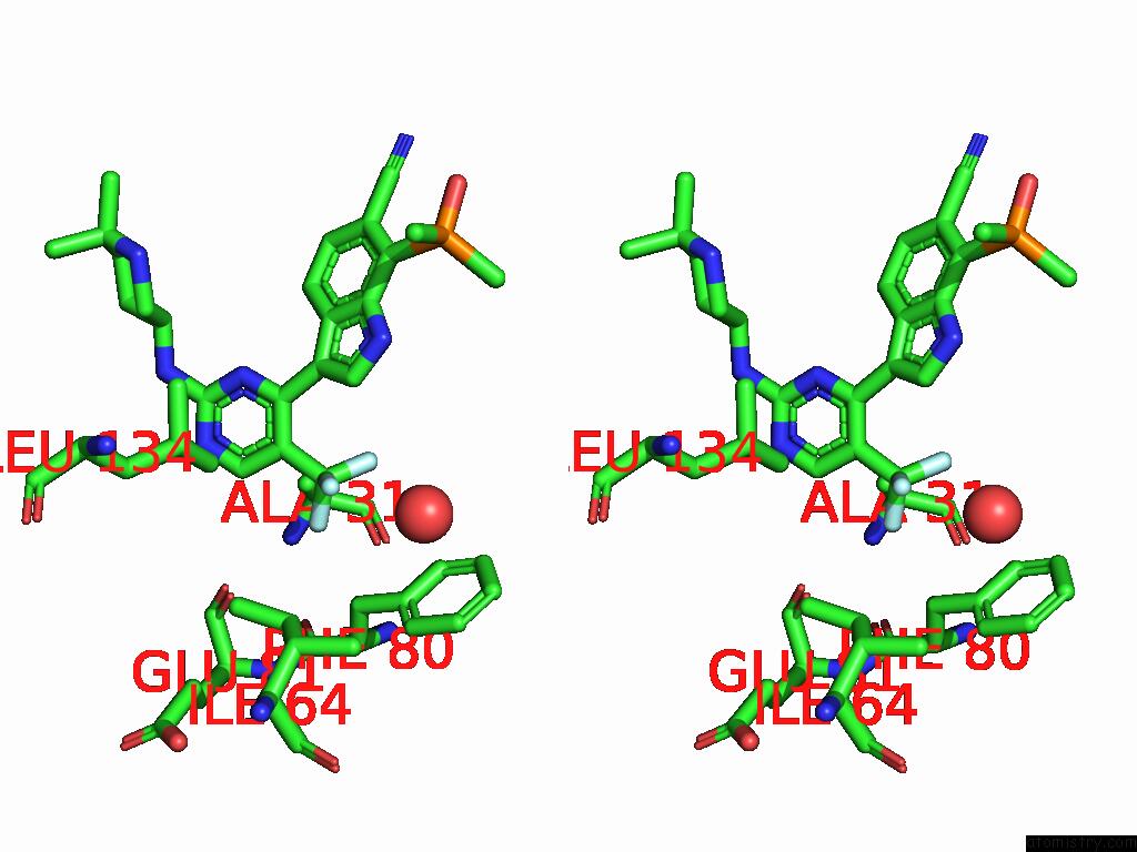
Fluorine binding site 1 out of 3 in 9gla
Go back to
Fluorine binding site 1 out
of 3 in the Crystal Structure of A CDK2-Based CDK7 Mimic with Inhibitor SY5609
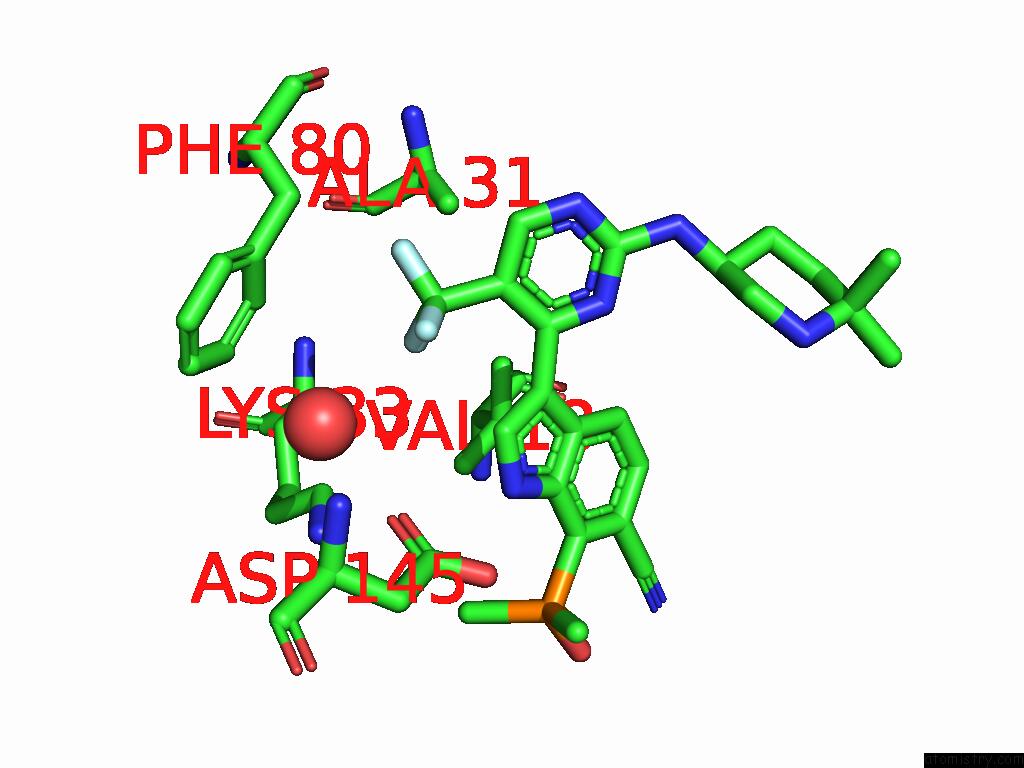
Mono view
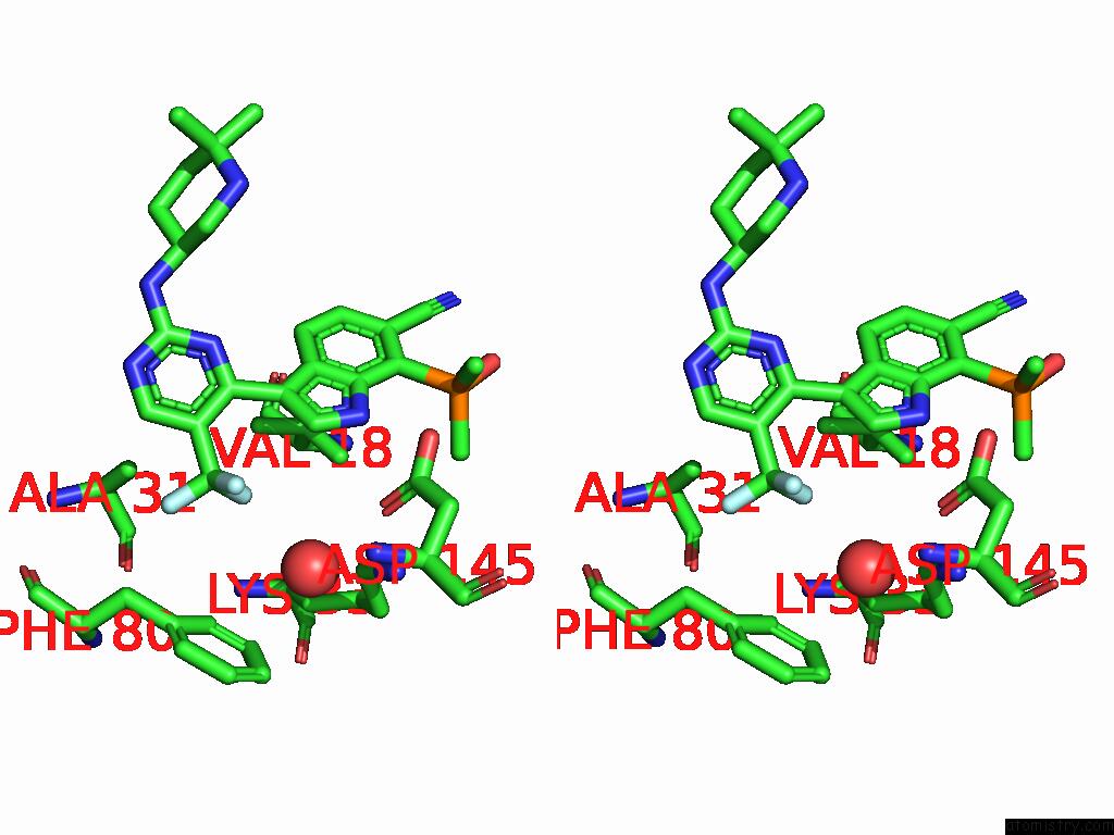
Stereo pair view
Mono view
Stereo pair view
A full contact list of Fluorine with other atoms in the F binding
site number 1 of Crystal Structure of A CDK2-Based CDK7 Mimic with Inhibitor SY5609 within 5.0Å range:
|
Fluorine binding site 2 out of 3 in 9gla
Go back to
Fluorine binding site 2 out
of 3 in the Crystal Structure of A CDK2-Based CDK7 Mimic with Inhibitor SY5609
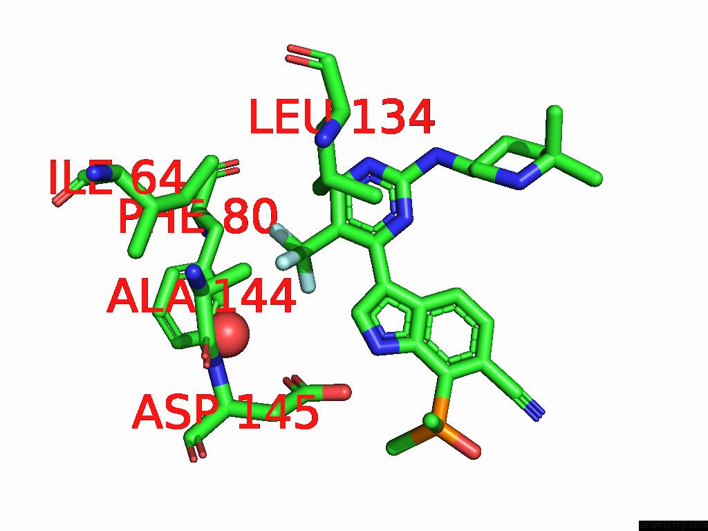
Mono view
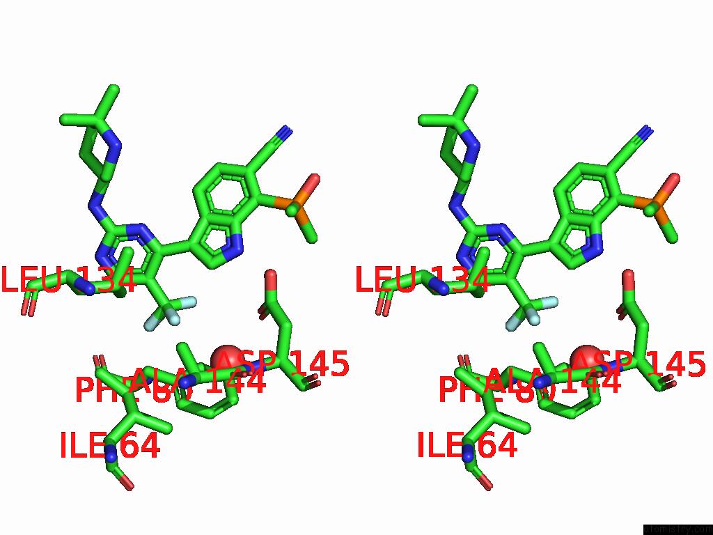
Stereo pair view
Mono view
Stereo pair view
A full contact list of Fluorine with other atoms in the F binding
site number 2 of Crystal Structure of A CDK2-Based CDK7 Mimic with Inhibitor SY5609 within 5.0Å range:
|
Fluorine binding site 3 out of 3 in 9gla
Go back to
Fluorine binding site 3 out
of 3 in the Crystal Structure of A CDK2-Based CDK7 Mimic with Inhibitor SY5609
Mono view
Stereo pair view
Mono view
Stereo pair view
A full contact list of Fluorine with other atoms in the F binding
site number 3 of Crystal Structure of A CDK2-Based CDK7 Mimic with Inhibitor SY5609 within 5.0Å range:
|
Reference:
J.Skerlova,
V.Krejcirikova,
M.Perina,
V.Vojackova,
M.Fabry,
V.Krystof,
R.Jorda,
P.Rezacova.
CDK2-Based CDK7 Mimic As A Tool For Structural Analysis: Biochemical Validation and Crystal Structure with SY5609. Int.J.Biol.Macromol. V. 294 39117 2025.
ISSN: ISSN 0141-8130
PubMed: 39733900
DOI: 10.1016/J.IJBIOMAC.2024.139117
Page generated: Wed Jul 16 11:39:26 2025
ISSN: ISSN 0141-8130
PubMed: 39733900
DOI: 10.1016/J.IJBIOMAC.2024.139117
Last articles
Fe in 7CX8Fe in 7CY4
Fe in 7CXZ
Fe in 7CPI
Fe in 7CUP
Fe in 7CUW
Fe in 7CUE
Fe in 7CUQ
Fe in 7CTC
Fe in 7CT7