Fluorine »
PDB 1bwf-1dvy »
1dvy »
Fluorine in PDB 1dvy: Crystal Structure of Transthyretin in Complex with N-(M- Trifluoromethylphenyl) Phenoxazine-4,6-Dicarboxylic Acid
Protein crystallography data
The structure of Crystal Structure of Transthyretin in Complex with N-(M- Trifluoromethylphenyl) Phenoxazine-4,6-Dicarboxylic Acid, PDB code: 1dvy
was solved by
T.Klabunde,
H.M.Petrassi,
V.B.Oza,
J.W.Kelly,
J.C.Sacchettini,
with X-Ray Crystallography technique. A brief refinement statistics is given in the table below:
| Resolution Low / High (Å) | 8.00 / 1.90 |
| Space group | P 21 21 2 |
| Cell size a, b, c (Å), α, β, γ (°) | 43.290, 86.030, 65.230, 90.00, 90.00, 90.00 |
| R / Rfree (%) | 20.2 / 20.3 |
Fluorine Binding Sites:
The binding sites of Fluorine atom in the Crystal Structure of Transthyretin in Complex with N-(M- Trifluoromethylphenyl) Phenoxazine-4,6-Dicarboxylic Acid
(pdb code 1dvy). This binding sites where shown within
5.0 Angstroms radius around Fluorine atom.
In total 6 binding sites of Fluorine where determined in the Crystal Structure of Transthyretin in Complex with N-(M- Trifluoromethylphenyl) Phenoxazine-4,6-Dicarboxylic Acid, PDB code: 1dvy:
Jump to Fluorine binding site number: 1; 2; 3; 4; 5; 6;
In total 6 binding sites of Fluorine where determined in the Crystal Structure of Transthyretin in Complex with N-(M- Trifluoromethylphenyl) Phenoxazine-4,6-Dicarboxylic Acid, PDB code: 1dvy:
Jump to Fluorine binding site number: 1; 2; 3; 4; 5; 6;
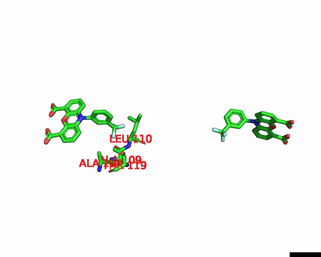
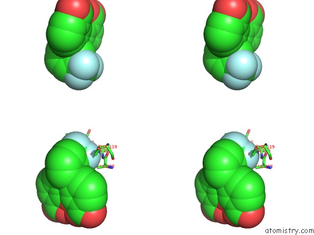
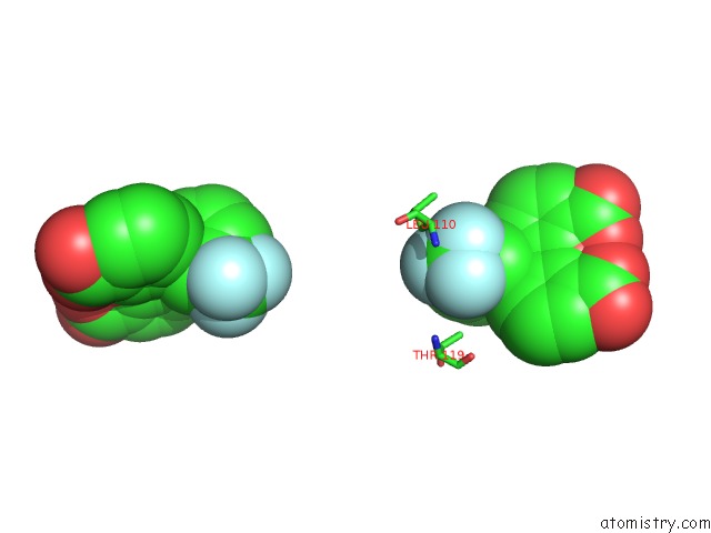
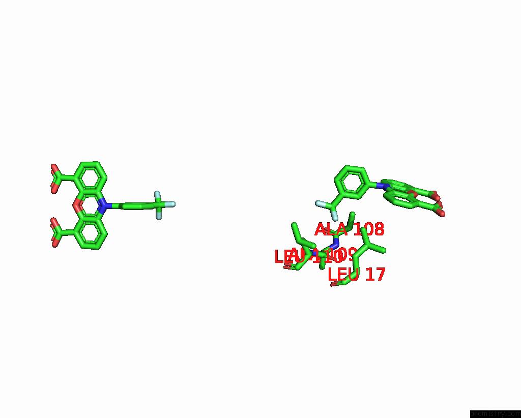
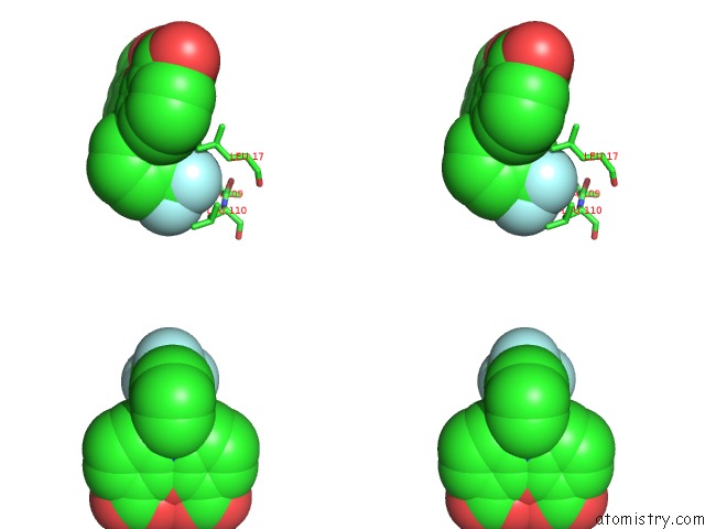
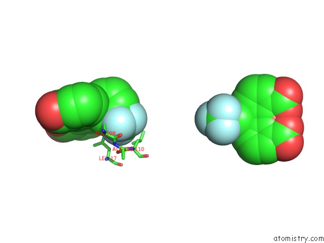
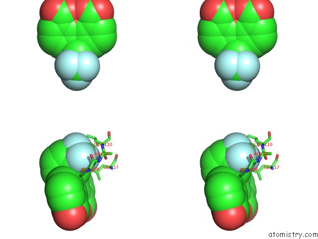
Fluorine binding site 1 out of 6 in 1dvy
Go back to
Fluorine binding site 1 out
of 6 in the Crystal Structure of Transthyretin in Complex with N-(M- Trifluoromethylphenyl) Phenoxazine-4,6-Dicarboxylic Acid
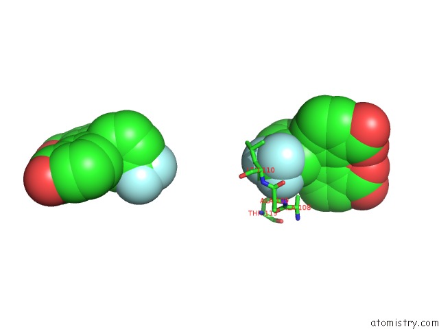
Mono view
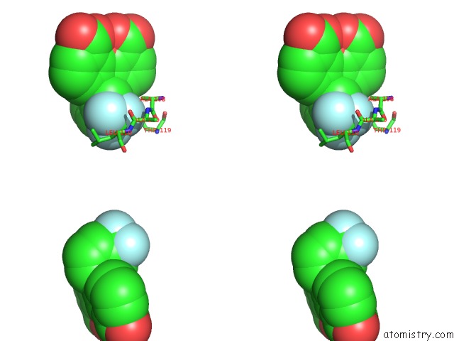
Stereo pair view
Mono view
Stereo pair view
A full contact list of Fluorine with other atoms in the F binding
site number 1 of Crystal Structure of Transthyretin in Complex with N-(M- Trifluoromethylphenyl) Phenoxazine-4,6-Dicarboxylic Acid within 5.0Å range:
|
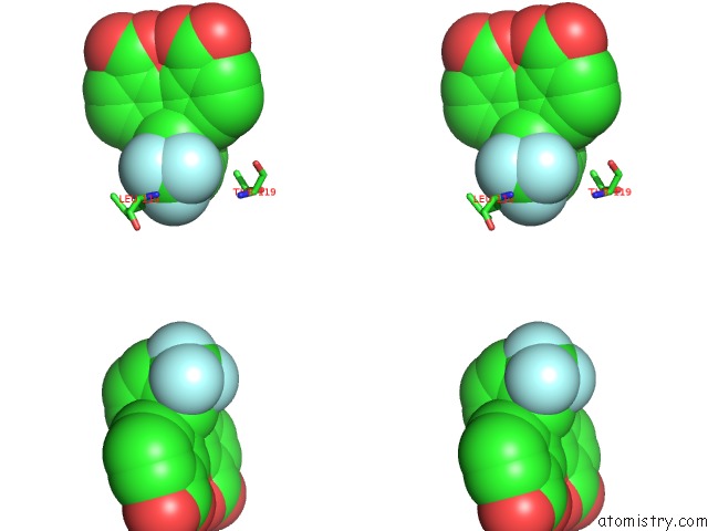
Fluorine binding site 2 out of 6 in 1dvy
Go back to
Fluorine binding site 2 out
of 6 in the Crystal Structure of Transthyretin in Complex with N-(M- Trifluoromethylphenyl) Phenoxazine-4,6-Dicarboxylic Acid
Mono view
Stereo pair view
Mono view
Stereo pair view
A full contact list of Fluorine with other atoms in the F binding
site number 2 of Crystal Structure of Transthyretin in Complex with N-(M- Trifluoromethylphenyl) Phenoxazine-4,6-Dicarboxylic Acid within 5.0Å range:
|
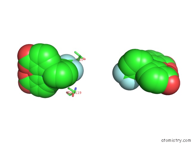
Fluorine binding site 3 out of 6 in 1dvy
Go back to
Fluorine binding site 3 out
of 6 in the Crystal Structure of Transthyretin in Complex with N-(M- Trifluoromethylphenyl) Phenoxazine-4,6-Dicarboxylic Acid
Mono view
Stereo pair view
Mono view
Stereo pair view
A full contact list of Fluorine with other atoms in the F binding
site number 3 of Crystal Structure of Transthyretin in Complex with N-(M- Trifluoromethylphenyl) Phenoxazine-4,6-Dicarboxylic Acid within 5.0Å range:
|
Fluorine binding site 4 out of 6 in 1dvy
Go back to
Fluorine binding site 4 out
of 6 in the Crystal Structure of Transthyretin in Complex with N-(M- Trifluoromethylphenyl) Phenoxazine-4,6-Dicarboxylic Acid
Mono view
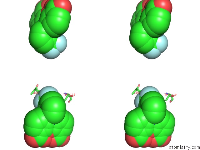
Stereo pair view
Mono view
Stereo pair view
A full contact list of Fluorine with other atoms in the F binding
site number 4 of Crystal Structure of Transthyretin in Complex with N-(M- Trifluoromethylphenyl) Phenoxazine-4,6-Dicarboxylic Acid within 5.0Å range:
|
Fluorine binding site 5 out of 6 in 1dvy
Go back to
Fluorine binding site 5 out
of 6 in the Crystal Structure of Transthyretin in Complex with N-(M- Trifluoromethylphenyl) Phenoxazine-4,6-Dicarboxylic Acid
Mono view
Stereo pair view
Mono view
Stereo pair view
A full contact list of Fluorine with other atoms in the F binding
site number 5 of Crystal Structure of Transthyretin in Complex with N-(M- Trifluoromethylphenyl) Phenoxazine-4,6-Dicarboxylic Acid within 5.0Å range:
|
Fluorine binding site 6 out of 6 in 1dvy
Go back to
Fluorine binding site 6 out
of 6 in the Crystal Structure of Transthyretin in Complex with N-(M- Trifluoromethylphenyl) Phenoxazine-4,6-Dicarboxylic Acid
Mono view
Stereo pair view
Mono view
Stereo pair view
A full contact list of Fluorine with other atoms in the F binding
site number 6 of Crystal Structure of Transthyretin in Complex with N-(M- Trifluoromethylphenyl) Phenoxazine-4,6-Dicarboxylic Acid within 5.0Å range:
|
Reference:
T.Klabunde,
H.M.Petrassi,
V.B.Oza,
P.Raman,
J.W.Kelly,
J.C.Sacchettini.
Rational Design of Potent Human Transthyretin Amyloid Disease Inhibitors. Nat.Struct.Biol. V. 7 312 2000.
ISSN: ISSN 1072-8368
PubMed: 10742177
DOI: 10.1038/74082
Page generated: Mon Jul 14 10:34:02 2025
ISSN: ISSN 1072-8368
PubMed: 10742177
DOI: 10.1038/74082
Last articles
Mg in 4JJSMg in 4JJ2
Mg in 4JIW
Mg in 4JIV
Mg in 4JIB
Mg in 4JI4
Mg in 4JI5
Mg in 4JI1
Mg in 4JI0
Mg in 4JI2