Fluorine »
PDB 1fko-1h1d »
1gw1 »
Fluorine in PDB 1gw1: Substrate Distortion By Beta-Mannanase From Pseudomonas Cellulosa
Enzymatic activity of Substrate Distortion By Beta-Mannanase From Pseudomonas Cellulosa
All present enzymatic activity of Substrate Distortion By Beta-Mannanase From Pseudomonas Cellulosa:
3.2.1.78;
3.2.1.78;
Protein crystallography data
The structure of Substrate Distortion By Beta-Mannanase From Pseudomonas Cellulosa, PDB code: 1gw1
was solved by
V.Ducros,
D.L.Zechel,
H.J.Gilbert,
L.Szabo,
S.G.Withers,
G.J.Davies,
with X-Ray Crystallography technique. A brief refinement statistics is given in the table below:
| Resolution Low / High (Å) | 20.00 / 1.65 |
| Space group | P 41 |
| Cell size a, b, c (Å), α, β, γ (°) | 93.488, 93.488, 53.719, 90.00, 90.00, 90.00 |
| R / Rfree (%) | 13.8 / 16.7 |
Other elements in 1gw1:
The structure of Substrate Distortion By Beta-Mannanase From Pseudomonas Cellulosa also contains other interesting chemical elements:
| Zinc | (Zn) | 2 atoms |
| Sodium | (Na) | 1 atom |
Fluorine Binding Sites:
The binding sites of Fluorine atom in the Substrate Distortion By Beta-Mannanase From Pseudomonas Cellulosa
(pdb code 1gw1). This binding sites where shown within
5.0 Angstroms radius around Fluorine atom.
In total 2 binding sites of Fluorine where determined in the Substrate Distortion By Beta-Mannanase From Pseudomonas Cellulosa, PDB code: 1gw1:
Jump to Fluorine binding site number: 1; 2;
In total 2 binding sites of Fluorine where determined in the Substrate Distortion By Beta-Mannanase From Pseudomonas Cellulosa, PDB code: 1gw1:
Jump to Fluorine binding site number: 1; 2;
Fluorine binding site 1 out of 2 in 1gw1
Go back to
Fluorine binding site 1 out
of 2 in the Substrate Distortion By Beta-Mannanase From Pseudomonas Cellulosa
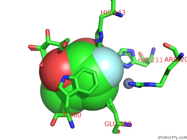
Mono view
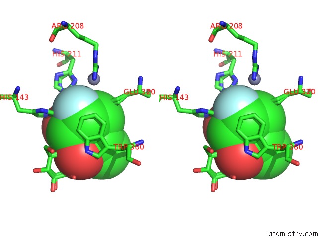
Stereo pair view
Mono view
Stereo pair view
A full contact list of Fluorine with other atoms in the F binding
site number 1 of Substrate Distortion By Beta-Mannanase From Pseudomonas Cellulosa within 5.0Å range:
|
Fluorine binding site 2 out of 2 in 1gw1
Go back to
Fluorine binding site 2 out
of 2 in the Substrate Distortion By Beta-Mannanase From Pseudomonas Cellulosa
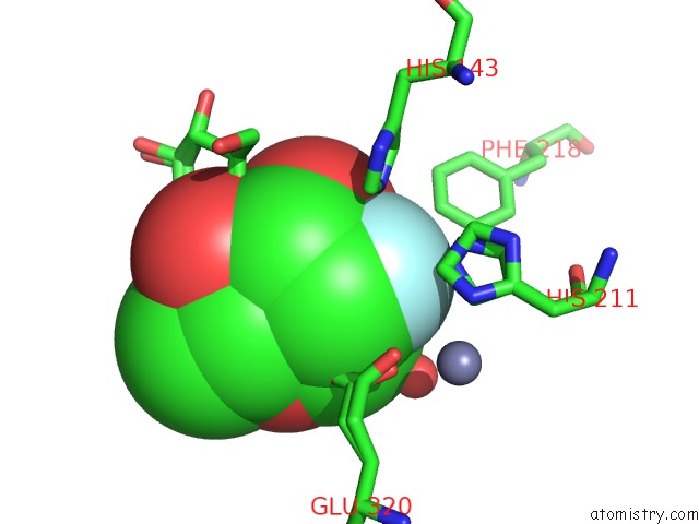
Mono view
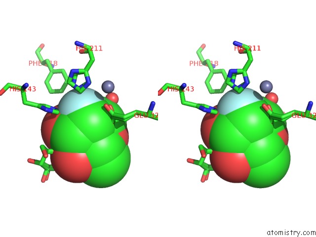
Stereo pair view
Mono view
Stereo pair view
A full contact list of Fluorine with other atoms in the F binding
site number 2 of Substrate Distortion By Beta-Mannanase From Pseudomonas Cellulosa within 5.0Å range:
|
Reference:
V.Ducros,
D.L.Zechel,
G.Murshudov,
H.J.Gilbert,
L.Szabo,
D.Stoll,
S.G.Withers,
G.J.Davies.
Substrate Distortion By A Beta-Mannanase: Snapshots of the Michaelis and Covalent-Intermediate Complexes Suggest A B2,5 Conformation For the Transition State Angew.Chem.Int.Ed.Engl. V. 41 2824 2002.
ISSN: ISSN 1433-7851
PubMed: 12203498
DOI: 10.1002/1521-3773(20020802)41:15<2824::AID-ANIE2824>3.0.CO;2
Page generated: Mon Jul 14 10:50:28 2025
ISSN: ISSN 1433-7851
PubMed: 12203498
DOI: 10.1002/1521-3773(20020802)41:15<2824::AID-ANIE2824>3.0.CO;2
Last articles
Fe in 6CKEFe in 6CIZ
Fe in 6CIR
Fe in 6CIQ
Fe in 6CDK
Fe in 6CII
Fe in 6CFW
Fe in 6CIF
Fe in 6CIE
Fe in 6CID