Fluorine »
PDB 1jdj-1mkd »
1k8w »
Fluorine in PDB 1k8w: Crystal Structure of the E. Coli Pseudouridine Synthase Trub Bound to A T Stem-Loop Rna
Enzymatic activity of Crystal Structure of the E. Coli Pseudouridine Synthase Trub Bound to A T Stem-Loop Rna
All present enzymatic activity of Crystal Structure of the E. Coli Pseudouridine Synthase Trub Bound to A T Stem-Loop Rna:
4.2.1.70;
4.2.1.70;
Protein crystallography data
The structure of Crystal Structure of the E. Coli Pseudouridine Synthase Trub Bound to A T Stem-Loop Rna, PDB code: 1k8w
was solved by
C.Hoang,
A.R.Ferre-D'amare,
with X-Ray Crystallography technique. A brief refinement statistics is given in the table below:
| Resolution Low / High (Å) | 28.27 / 1.85 |
| Space group | C 1 2 1 |
| Cell size a, b, c (Å), α, β, γ (°) | 145.054, 40.361, 77.987, 90.00, 110.60, 90.00 |
| R / Rfree (%) | 18.4 / 21.1 |
Fluorine Binding Sites:
The binding sites of Fluorine atom in the Crystal Structure of the E. Coli Pseudouridine Synthase Trub Bound to A T Stem-Loop Rna
(pdb code 1k8w). This binding sites where shown within
5.0 Angstroms radius around Fluorine atom.
In total only one binding site of Fluorine was determined in the Crystal Structure of the E. Coli Pseudouridine Synthase Trub Bound to A T Stem-Loop Rna, PDB code: 1k8w:
In total only one binding site of Fluorine was determined in the Crystal Structure of the E. Coli Pseudouridine Synthase Trub Bound to A T Stem-Loop Rna, PDB code: 1k8w:
Fluorine binding site 1 out of 1 in 1k8w
Go back to
Fluorine binding site 1 out
of 1 in the Crystal Structure of the E. Coli Pseudouridine Synthase Trub Bound to A T Stem-Loop Rna
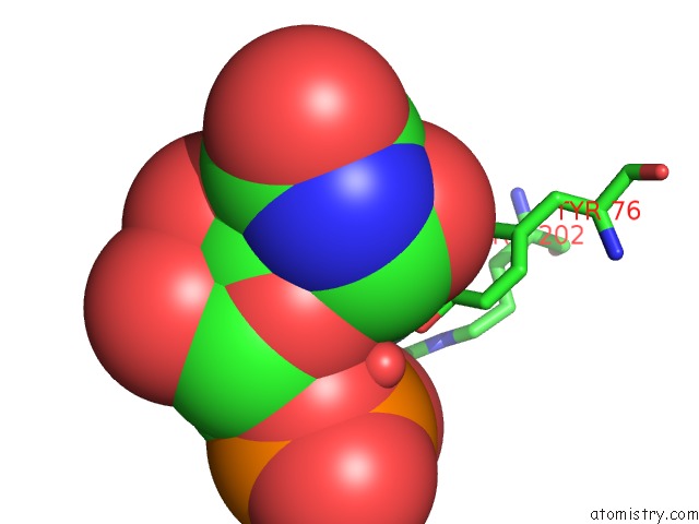
Mono view
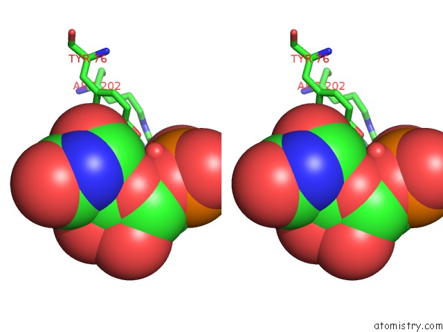
Stereo pair view
Mono view
Stereo pair view
A full contact list of Fluorine with other atoms in the F binding
site number 1 of Crystal Structure of the E. Coli Pseudouridine Synthase Trub Bound to A T Stem-Loop Rna within 5.0Å range:
|
Reference:
C.Hoang,
A.R.Ferre-D'amare.
Cocrystal Structure of A Trna PSI55 Pseudouridine Synthase: Nucleotide Flipping By An Rna-Modifying Enzyme. Cell(Cambridge,Mass.) V. 107 929 2001.
ISSN: ISSN 0092-8674
PubMed: 11779468
DOI: 10.1016/S0092-8674(01)00618-3
Page generated: Mon Jul 14 11:02:06 2025
ISSN: ISSN 0092-8674
PubMed: 11779468
DOI: 10.1016/S0092-8674(01)00618-3
Last articles
Mg in 2HJOMg in 2HIY
Mg in 2HHV
Mg in 2HHK
Mg in 2HHU
Mg in 2HHT
Mg in 2HH9
Mg in 2HHS
Mg in 2HHQ
Mg in 2HHP