Fluorine »
PDB 1mmd-1o5f »
1nhx »
Fluorine in PDB 1nhx: Pepck Complex with A Gtp-Competitive Inhibitor
Enzymatic activity of Pepck Complex with A Gtp-Competitive Inhibitor
All present enzymatic activity of Pepck Complex with A Gtp-Competitive Inhibitor:
4.1.1.32;
4.1.1.32;
Protein crystallography data
The structure of Pepck Complex with A Gtp-Competitive Inhibitor, PDB code: 1nhx
was solved by
L.H.Foley,
P.Wang,
P.Dunten,
G.Ramsey,
M.-L.Gubler,
S.J.Wertheimer,
with X-Ray Crystallography technique. A brief refinement statistics is given in the table below:
| Resolution Low / High (Å) | 50.00 / 2.10 |
| Space group | P 1 |
| Cell size a, b, c (Å), α, β, γ (°) | 45.383, 61.452, 62.265, 89.71, 70.24, 72.56 |
| R / Rfree (%) | 17.3 / 21.7 |
Other elements in 1nhx:
The structure of Pepck Complex with A Gtp-Competitive Inhibitor also contains other interesting chemical elements:
| Manganese | (Mn) | 1 atom |
| Sodium | (Na) | 1 atom |
Fluorine Binding Sites:
The binding sites of Fluorine atom in the Pepck Complex with A Gtp-Competitive Inhibitor
(pdb code 1nhx). This binding sites where shown within
5.0 Angstroms radius around Fluorine atom.
In total only one binding site of Fluorine was determined in the Pepck Complex with A Gtp-Competitive Inhibitor, PDB code: 1nhx:
In total only one binding site of Fluorine was determined in the Pepck Complex with A Gtp-Competitive Inhibitor, PDB code: 1nhx:
Fluorine binding site 1 out of 1 in 1nhx
Go back to
Fluorine binding site 1 out
of 1 in the Pepck Complex with A Gtp-Competitive Inhibitor
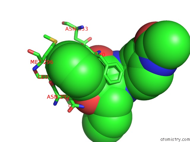
Mono view
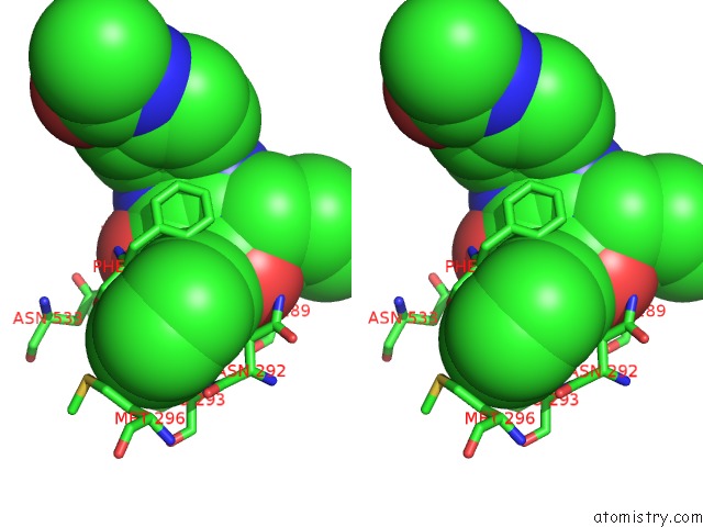
Stereo pair view
Mono view
Stereo pair view
A full contact list of Fluorine with other atoms in the F binding
site number 1 of Pepck Complex with A Gtp-Competitive Inhibitor within 5.0Å range:
|
Reference:
L.H.Foley,
P.Wang,
P.Dunten,
G.Ramsey,
M.-L.Gubler,
S.J.Wertheimer.
X-Ray Structures of Two Xanthine Inhibitors Bound to Pepck and N-3 Modifications of Substituted 1,8-Dibenzylxanthines Bioorg.Med.Chem.Lett. V. 13 3871 2003.
ISSN: ISSN 0960-894X
PubMed: 14552798
DOI: 10.1016/S0960-894X(03)00723-6
Page generated: Mon Jul 14 11:22:01 2025
ISSN: ISSN 0960-894X
PubMed: 14552798
DOI: 10.1016/S0960-894X(03)00723-6
Last articles
K in 4RN2K in 4RN1
K in 4RP3
K in 4RN0
K in 4RLG
K in 4RGQ
K in 4RKJ
K in 4RJ9
K in 4RJZ
K in 4RES