Fluorine »
PDB 1rw8-1uda »
1ta0 »
Fluorine in PDB 1ta0: Three-Dimensional Structure of A Rna-Polymerase II Binding Protein with Associated Ligand.
Enzymatic activity of Three-Dimensional Structure of A Rna-Polymerase II Binding Protein with Associated Ligand.
All present enzymatic activity of Three-Dimensional Structure of A Rna-Polymerase II Binding Protein with Associated Ligand.:
3.1.3.16;
3.1.3.16;
Protein crystallography data
The structure of Three-Dimensional Structure of A Rna-Polymerase II Binding Protein with Associated Ligand., PDB code: 1ta0
was solved by
T.Kamenski,
S.Heilmeier,
T.Meinhart,
P.Cramer,
with X-Ray Crystallography technique. A brief refinement statistics is given in the table below:
| Resolution Low / High (Å) | 20.00 / 2.10 |
| Space group | P 21 21 2 |
| Cell size a, b, c (Å), α, β, γ (°) | 117.820, 47.170, 40.070, 90.00, 90.00, 90.00 |
| R / Rfree (%) | 20.7 / 22.7 |
Other elements in 1ta0:
The structure of Three-Dimensional Structure of A Rna-Polymerase II Binding Protein with Associated Ligand. also contains other interesting chemical elements:
| Magnesium | (Mg) | 1 atom |
Fluorine Binding Sites:
The binding sites of Fluorine atom in the Three-Dimensional Structure of A Rna-Polymerase II Binding Protein with Associated Ligand.
(pdb code 1ta0). This binding sites where shown within
5.0 Angstroms radius around Fluorine atom.
In total 3 binding sites of Fluorine where determined in the Three-Dimensional Structure of A Rna-Polymerase II Binding Protein with Associated Ligand., PDB code: 1ta0:
Jump to Fluorine binding site number: 1; 2; 3;
In total 3 binding sites of Fluorine where determined in the Three-Dimensional Structure of A Rna-Polymerase II Binding Protein with Associated Ligand., PDB code: 1ta0:
Jump to Fluorine binding site number: 1; 2; 3;
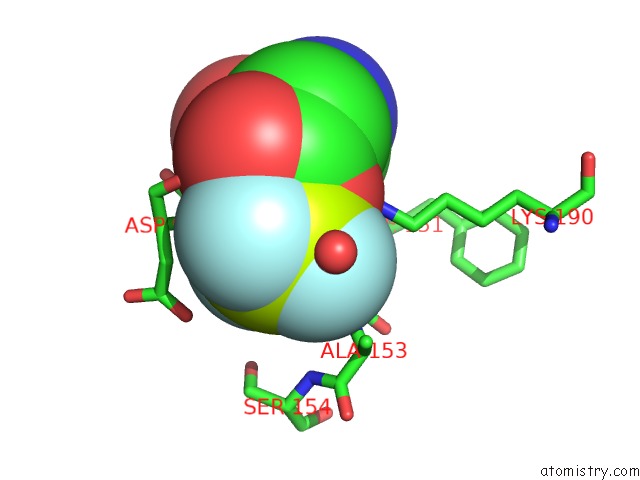
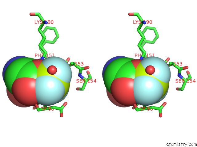
Fluorine binding site 1 out of 3 in 1ta0
Go back to
Fluorine binding site 1 out
of 3 in the Three-Dimensional Structure of A Rna-Polymerase II Binding Protein with Associated Ligand.
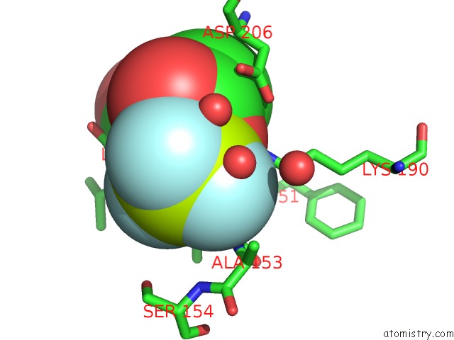
Mono view
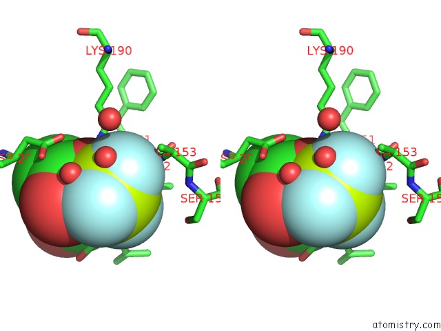
Stereo pair view
Mono view
Stereo pair view
A full contact list of Fluorine with other atoms in the F binding
site number 1 of Three-Dimensional Structure of A Rna-Polymerase II Binding Protein with Associated Ligand. within 5.0Å range:
|
Fluorine binding site 2 out of 3 in 1ta0
Go back to
Fluorine binding site 2 out
of 3 in the Three-Dimensional Structure of A Rna-Polymerase II Binding Protein with Associated Ligand.
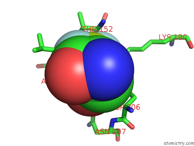
Mono view
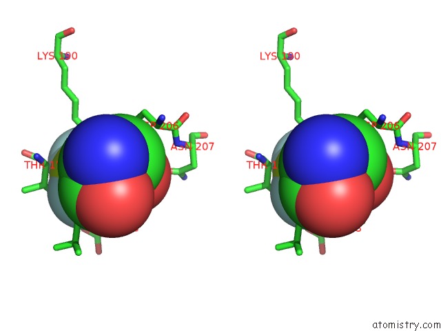
Stereo pair view
Mono view
Stereo pair view
A full contact list of Fluorine with other atoms in the F binding
site number 2 of Three-Dimensional Structure of A Rna-Polymerase II Binding Protein with Associated Ligand. within 5.0Å range:
|
Fluorine binding site 3 out of 3 in 1ta0
Go back to
Fluorine binding site 3 out
of 3 in the Three-Dimensional Structure of A Rna-Polymerase II Binding Protein with Associated Ligand.
Mono view
Stereo pair view
Mono view
Stereo pair view
A full contact list of Fluorine with other atoms in the F binding
site number 3 of Three-Dimensional Structure of A Rna-Polymerase II Binding Protein with Associated Ligand. within 5.0Å range:
|
Reference:
T.Kamenski,
S.Heilmeier,
T.Meinhart,
P.Cramer.
Structure and Mechanism of Rna Polymerase II Ctd Phosphatases. Mol.Cell V. 15 399 2004.
ISSN: ISSN 1097-2765
PubMed: 15304220
DOI: 10.1016/J.MOLCEL.2004.06.035
Page generated: Mon Jul 14 11:59:32 2025
ISSN: ISSN 1097-2765
PubMed: 15304220
DOI: 10.1016/J.MOLCEL.2004.06.035
Last articles
K in 7KURK in 7KUQ
K in 7KLU
K in 7KLP
K in 7KNC
K in 7KLV
K in 7KMP
K in 7KDY
K in 7KLB
K in 7KD9