Fluorine »
PDB 1udb-1w5y »
1upv »
Fluorine in PDB 1upv: Crystal Structure of the Human Liver X Receptor Beta Ligand Binding Domain in Complex with A Synthetic Agonist
Protein crystallography data
The structure of Crystal Structure of the Human Liver X Receptor Beta Ligand Binding Domain in Complex with A Synthetic Agonist, PDB code: 1upv
was solved by
S.Hoerer,
A.Schmid,
A.Heckel,
R.M.Budzinski,
H.Nar,
with X-Ray Crystallography technique. A brief refinement statistics is given in the table below:
| Resolution Low / High (Å) | 20.00 / 2.10 |
| Space group | C 1 2 1 |
| Cell size a, b, c (Å), α, β, γ (°) | 141.800, 43.200, 51.800, 90.00, 105.80, 90.00 |
| R / Rfree (%) | 21.5 / 27.4 |
Fluorine Binding Sites:
The binding sites of Fluorine atom in the Crystal Structure of the Human Liver X Receptor Beta Ligand Binding Domain in Complex with A Synthetic Agonist
(pdb code 1upv). This binding sites where shown within
5.0 Angstroms radius around Fluorine atom.
In total 9 binding sites of Fluorine where determined in the Crystal Structure of the Human Liver X Receptor Beta Ligand Binding Domain in Complex with A Synthetic Agonist, PDB code: 1upv:
Jump to Fluorine binding site number: 1; 2; 3; 4; 5; 6; 7; 8; 9;
In total 9 binding sites of Fluorine where determined in the Crystal Structure of the Human Liver X Receptor Beta Ligand Binding Domain in Complex with A Synthetic Agonist, PDB code: 1upv:
Jump to Fluorine binding site number: 1; 2; 3; 4; 5; 6; 7; 8; 9;
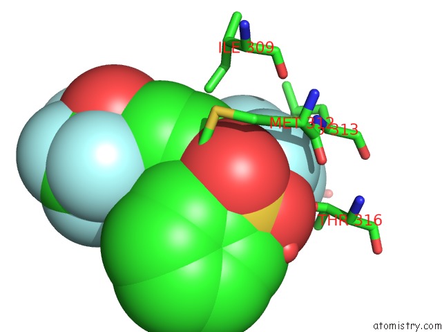
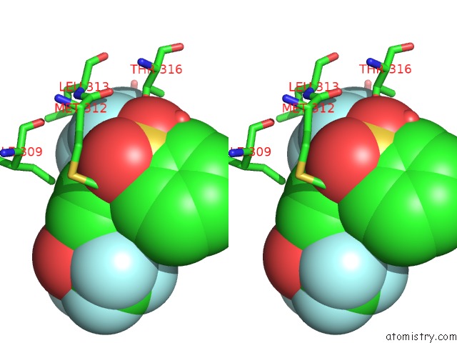
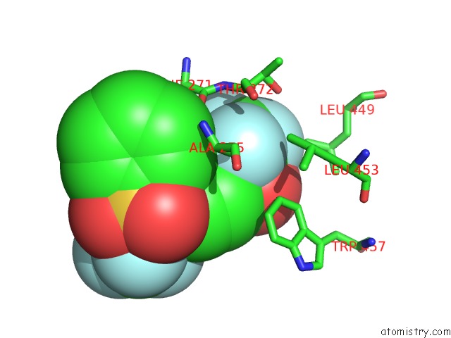
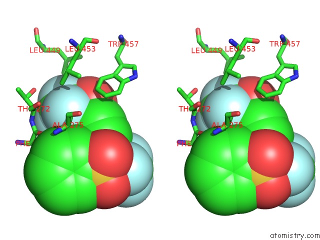
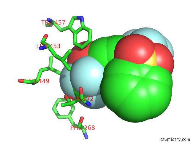
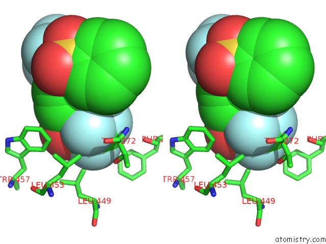
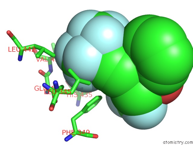
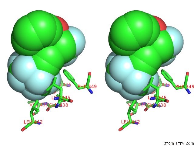
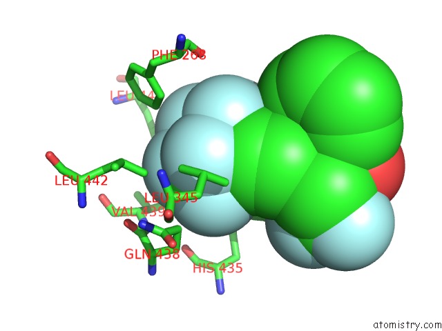
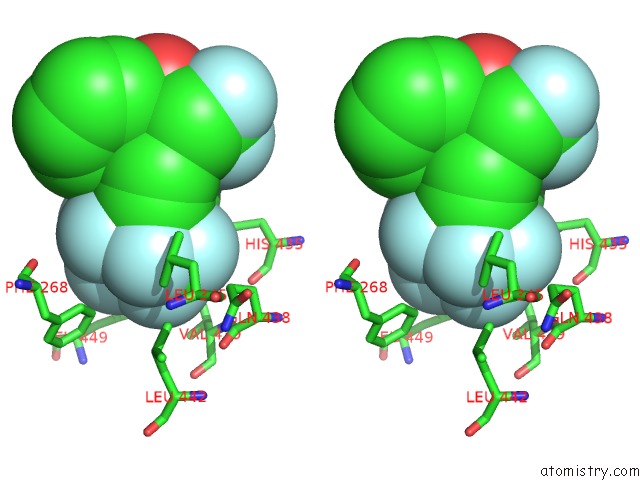
Fluorine binding site 1 out of 9 in 1upv
Go back to
Fluorine binding site 1 out
of 9 in the Crystal Structure of the Human Liver X Receptor Beta Ligand Binding Domain in Complex with A Synthetic Agonist
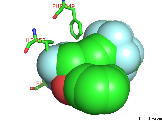
Mono view
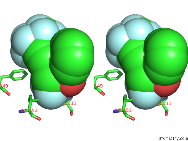
Stereo pair view
Mono view
Stereo pair view
A full contact list of Fluorine with other atoms in the F binding
site number 1 of Crystal Structure of the Human Liver X Receptor Beta Ligand Binding Domain in Complex with A Synthetic Agonist within 5.0Å range:
|
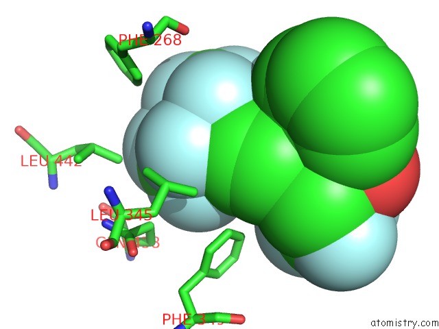
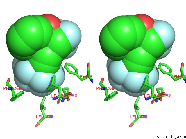
Fluorine binding site 2 out of 9 in 1upv
Go back to
Fluorine binding site 2 out
of 9 in the Crystal Structure of the Human Liver X Receptor Beta Ligand Binding Domain in Complex with A Synthetic Agonist
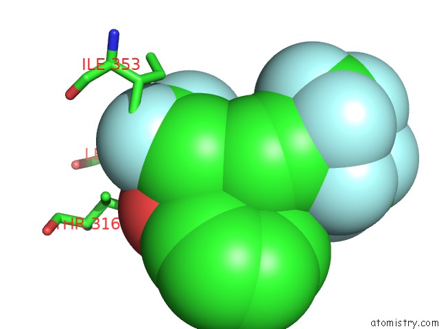
Mono view
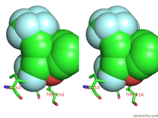
Stereo pair view
Mono view
Stereo pair view
A full contact list of Fluorine with other atoms in the F binding
site number 2 of Crystal Structure of the Human Liver X Receptor Beta Ligand Binding Domain in Complex with A Synthetic Agonist within 5.0Å range:
|
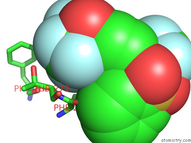
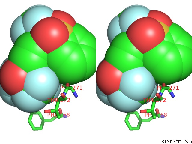
Fluorine binding site 3 out of 9 in 1upv
Go back to
Fluorine binding site 3 out
of 9 in the Crystal Structure of the Human Liver X Receptor Beta Ligand Binding Domain in Complex with A Synthetic Agonist
Mono view
Stereo pair view
Mono view
Stereo pair view
A full contact list of Fluorine with other atoms in the F binding
site number 3 of Crystal Structure of the Human Liver X Receptor Beta Ligand Binding Domain in Complex with A Synthetic Agonist within 5.0Å range:
|
Fluorine binding site 4 out of 9 in 1upv
Go back to
Fluorine binding site 4 out
of 9 in the Crystal Structure of the Human Liver X Receptor Beta Ligand Binding Domain in Complex with A Synthetic Agonist
Mono view
Stereo pair view
Mono view
Stereo pair view
A full contact list of Fluorine with other atoms in the F binding
site number 4 of Crystal Structure of the Human Liver X Receptor Beta Ligand Binding Domain in Complex with A Synthetic Agonist within 5.0Å range:
|
Fluorine binding site 5 out of 9 in 1upv
Go back to
Fluorine binding site 5 out
of 9 in the Crystal Structure of the Human Liver X Receptor Beta Ligand Binding Domain in Complex with A Synthetic Agonist
Mono view
Stereo pair view
Mono view
Stereo pair view
A full contact list of Fluorine with other atoms in the F binding
site number 5 of Crystal Structure of the Human Liver X Receptor Beta Ligand Binding Domain in Complex with A Synthetic Agonist within 5.0Å range:
|
Fluorine binding site 6 out of 9 in 1upv
Go back to
Fluorine binding site 6 out
of 9 in the Crystal Structure of the Human Liver X Receptor Beta Ligand Binding Domain in Complex with A Synthetic Agonist
Mono view
Stereo pair view
Mono view
Stereo pair view
A full contact list of Fluorine with other atoms in the F binding
site number 6 of Crystal Structure of the Human Liver X Receptor Beta Ligand Binding Domain in Complex with A Synthetic Agonist within 5.0Å range:
|
Fluorine binding site 7 out of 9 in 1upv
Go back to
Fluorine binding site 7 out
of 9 in the Crystal Structure of the Human Liver X Receptor Beta Ligand Binding Domain in Complex with A Synthetic Agonist
Mono view
Stereo pair view
Mono view
Stereo pair view
A full contact list of Fluorine with other atoms in the F binding
site number 7 of Crystal Structure of the Human Liver X Receptor Beta Ligand Binding Domain in Complex with A Synthetic Agonist within 5.0Å range:
|
Fluorine binding site 8 out of 9 in 1upv
Go back to
Fluorine binding site 8 out
of 9 in the Crystal Structure of the Human Liver X Receptor Beta Ligand Binding Domain in Complex with A Synthetic Agonist
Mono view
Stereo pair view
Mono view
Stereo pair view
A full contact list of Fluorine with other atoms in the F binding
site number 8 of Crystal Structure of the Human Liver X Receptor Beta Ligand Binding Domain in Complex with A Synthetic Agonist within 5.0Å range:
|
Fluorine binding site 9 out of 9 in 1upv
Go back to
Fluorine binding site 9 out
of 9 in the Crystal Structure of the Human Liver X Receptor Beta Ligand Binding Domain in Complex with A Synthetic Agonist
Mono view
Stereo pair view
Mono view
Stereo pair view
A full contact list of Fluorine with other atoms in the F binding
site number 9 of Crystal Structure of the Human Liver X Receptor Beta Ligand Binding Domain in Complex with A Synthetic Agonist within 5.0Å range:
|
Reference:
S.Hoerer,
A.Schmid,
A.Heckel,
R.M.Budzinski,
H.Nar.
Crystal Structure of the Human Liver X Receptor Beta Ligand-Binding Domain in Complex with A Synthetic Agonist J.Mol.Biol. V. 334 853 2003.
ISSN: ISSN 0022-2836
PubMed: 14643652
DOI: 10.1016/J.JMB.2003.10.033
Page generated: Mon Jul 14 12:03:34 2025
ISSN: ISSN 0022-2836
PubMed: 14643652
DOI: 10.1016/J.JMB.2003.10.033
Last articles
Fe in 7BI7Fe in 7BHC
Fe in 7BIU
Fe in 7BI1
Fe in 7B9A
Fe in 7BGI
Fe in 7BHB
Fe in 7B97
Fe in 7BHA
Fe in 7BBT