Fluorine »
PDB 2a4z-2baq »
2ax6 »
Fluorine in PDB 2ax6: Crystal Structure of the Androgen Receptor Ligand Binding Domain T877A Mutant in Complex with Hydroxyflutamide
Protein crystallography data
The structure of Crystal Structure of the Androgen Receptor Ligand Binding Domain T877A Mutant in Complex with Hydroxyflutamide, PDB code: 2ax6
was solved by
C.E.Bohl,
D.D.Miller,
J.Chen,
C.E.Bell,
J.T.Dalton,
with X-Ray Crystallography technique. A brief refinement statistics is given in the table below:
| Resolution Low / High (Å) | 18.03 / 1.50 |
| Space group | P 21 21 21 |
| Cell size a, b, c (Å), α, β, γ (°) | 54.782, 65.965, 69.628, 90.00, 90.00, 90.00 |
| R / Rfree (%) | 24.6 / 27.4 |
Fluorine Binding Sites:
The binding sites of Fluorine atom in the Crystal Structure of the Androgen Receptor Ligand Binding Domain T877A Mutant in Complex with Hydroxyflutamide
(pdb code 2ax6). This binding sites where shown within
5.0 Angstroms radius around Fluorine atom.
In total 3 binding sites of Fluorine where determined in the Crystal Structure of the Androgen Receptor Ligand Binding Domain T877A Mutant in Complex with Hydroxyflutamide, PDB code: 2ax6:
Jump to Fluorine binding site number: 1; 2; 3;
In total 3 binding sites of Fluorine where determined in the Crystal Structure of the Androgen Receptor Ligand Binding Domain T877A Mutant in Complex with Hydroxyflutamide, PDB code: 2ax6:
Jump to Fluorine binding site number: 1; 2; 3;
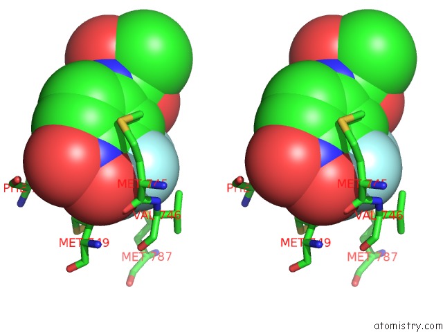
Fluorine binding site 1 out of 3 in 2ax6
Go back to
Fluorine binding site 1 out
of 3 in the Crystal Structure of the Androgen Receptor Ligand Binding Domain T877A Mutant in Complex with Hydroxyflutamide
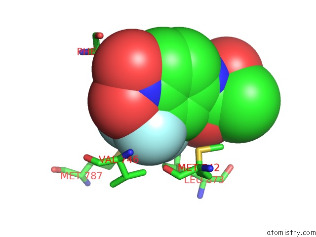
Mono view

Stereo pair view
Mono view
Stereo pair view
A full contact list of Fluorine with other atoms in the F binding
site number 1 of Crystal Structure of the Androgen Receptor Ligand Binding Domain T877A Mutant in Complex with Hydroxyflutamide within 5.0Å range:
|
Fluorine binding site 2 out of 3 in 2ax6
Go back to
Fluorine binding site 2 out
of 3 in the Crystal Structure of the Androgen Receptor Ligand Binding Domain T877A Mutant in Complex with Hydroxyflutamide
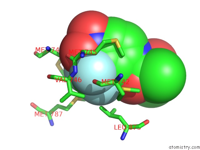
Mono view
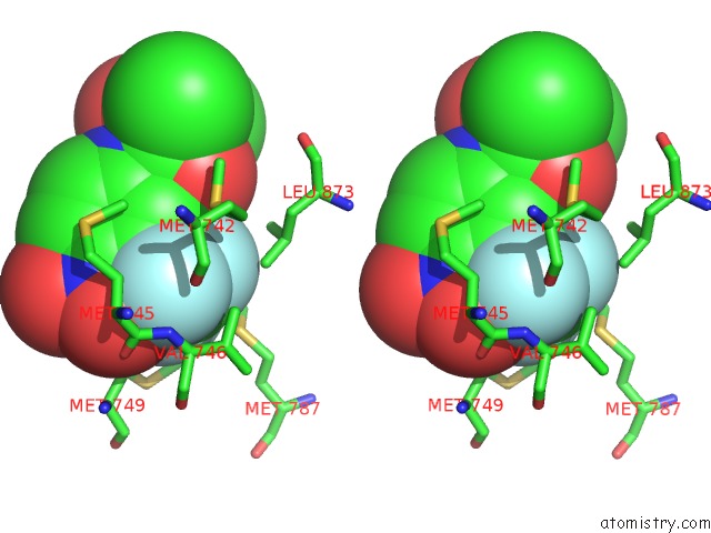
Stereo pair view
Mono view
Stereo pair view
A full contact list of Fluorine with other atoms in the F binding
site number 2 of Crystal Structure of the Androgen Receptor Ligand Binding Domain T877A Mutant in Complex with Hydroxyflutamide within 5.0Å range:
|
Fluorine binding site 3 out of 3 in 2ax6
Go back to
Fluorine binding site 3 out
of 3 in the Crystal Structure of the Androgen Receptor Ligand Binding Domain T877A Mutant in Complex with Hydroxyflutamide
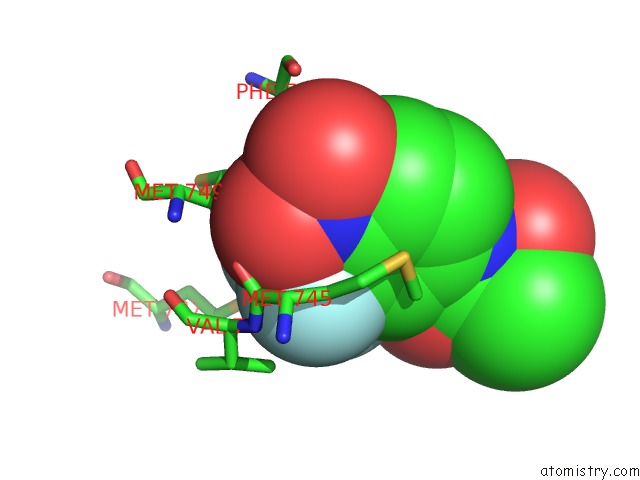
Mono view
Stereo pair view
Mono view
Stereo pair view
A full contact list of Fluorine with other atoms in the F binding
site number 3 of Crystal Structure of the Androgen Receptor Ligand Binding Domain T877A Mutant in Complex with Hydroxyflutamide within 5.0Å range:
|
Reference:
C.E.Bohl,
D.D.Miller,
J.Chen,
C.E.Bell,
J.T.Dalton.
Structural Basis For Accommodation of Nonsteroidal Ligands in the Androgen Receptor J.Biol.Chem. V. 280 37747 2005.
ISSN: ISSN 0021-9258
PubMed: 16129672
DOI: 10.1074/JBC.M507464200
Page generated: Mon Jul 14 12:37:51 2025
ISSN: ISSN 0021-9258
PubMed: 16129672
DOI: 10.1074/JBC.M507464200
Last articles
Mg in 3T2CMg in 3T2B
Mg in 3T1R
Mg in 3T1O
Mg in 3T0D
Mg in 3T1Q
Mg in 3T12
Mg in 3T1K
Mg in 3T10
Mg in 3T0Z