Fluorine »
PDB 2k1q-2ogz »
2lh5 »
Fluorine in PDB 2lh5: X-Ray Structural Investigation of Leghemoglobin. VI. Structure of Acetate-Ferrileghemoglobin at A Resolution of 2.0 Angstroms (Russian)
Protein crystallography data
The structure of X-Ray Structural Investigation of Leghemoglobin. VI. Structure of Acetate-Ferrileghemoglobin at A Resolution of 2.0 Angstroms (Russian), PDB code: 2lh5
was solved by
B.K.Vainshtein,
E.H.Harutyunyan,
I.P.Kuranova,
V.V.Borisov,
N.I.Sosfenov,
A.G.Pavlovsky,
A.I.Grebenko,
N.V.Konareva,
with X-Ray Crystallography technique. A brief refinement statistics is given in the table below:
| Resolution Low / High (Å) | N/A / 2.00 |
| Space group | B 2 |
| Cell size a, b, c (Å), α, β, γ (°) | 93.340, 38.240, 51.910, 90.00, 90.00, 98.80 |
| R / Rfree (%) | n/a / n/a |
Other elements in 2lh5:
The structure of X-Ray Structural Investigation of Leghemoglobin. VI. Structure of Acetate-Ferrileghemoglobin at A Resolution of 2.0 Angstroms (Russian) also contains other interesting chemical elements:
| Iron | (Fe) | 1 atom |
Fluorine Binding Sites:
The binding sites of Fluorine atom in the X-Ray Structural Investigation of Leghemoglobin. VI. Structure of Acetate-Ferrileghemoglobin at A Resolution of 2.0 Angstroms (Russian)
(pdb code 2lh5). This binding sites where shown within
5.0 Angstroms radius around Fluorine atom.
In total only one binding site of Fluorine was determined in the X-Ray Structural Investigation of Leghemoglobin. VI. Structure of Acetate-Ferrileghemoglobin at A Resolution of 2.0 Angstroms (Russian), PDB code: 2lh5:
In total only one binding site of Fluorine was determined in the X-Ray Structural Investigation of Leghemoglobin. VI. Structure of Acetate-Ferrileghemoglobin at A Resolution of 2.0 Angstroms (Russian), PDB code: 2lh5:
Fluorine binding site 1 out of 1 in 2lh5
Go back to
Fluorine binding site 1 out
of 1 in the X-Ray Structural Investigation of Leghemoglobin. VI. Structure of Acetate-Ferrileghemoglobin at A Resolution of 2.0 Angstroms (Russian)
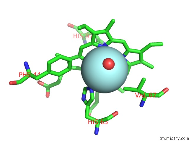
Mono view
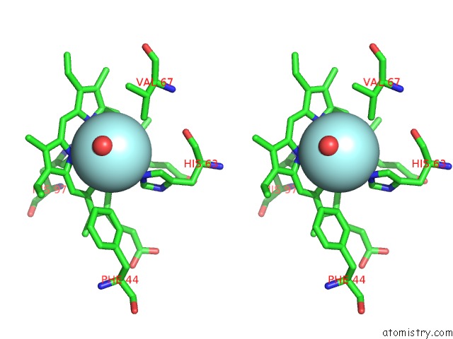
Stereo pair view
Mono view
Stereo pair view
A full contact list of Fluorine with other atoms in the F binding
site number 1 of X-Ray Structural Investigation of Leghemoglobin. VI. Structure of Acetate-Ferrileghemoglobin at A Resolution of 2.0 Angstroms (Russian) within 5.0Å range:
|
Reference:
E.G.Arutyunyan,
I.P.Kuranova,
B.K.Vainshtein,
W.Steigemann.
X-Ray Structural Investigation of Leghemoglobin. VI. Structure of Acetate-Ferrileghemoglobin at A Resolution of 2.0 Angstroms (Russian) Kristallografiya V. 25 80 1980.
ISSN: ISSN 0023-4761
Page generated: Mon Jul 14 13:38:39 2025
ISSN: ISSN 0023-4761
Last articles
Mg in 7DU2Mg in 7DSP
Mg in 7DSJ
Mg in 7DSI
Mg in 7DRP
Mg in 7DSH
Mg in 7DSA
Mg in 7DRX
Mg in 7DRM
Mg in 7DPT