Fluorine »
PDB 2rfn-2vfz »
2rhw »
Fluorine in PDB 2rhw: Crystal Structure of the S112A Mutant of A C-C Hydrolase, Bphd From Burkholderia Xenovorans LB400, in Complex with 3,10-Di-Fluoro Hopda
Protein crystallography data
The structure of Crystal Structure of the S112A Mutant of A C-C Hydrolase, Bphd From Burkholderia Xenovorans LB400, in Complex with 3,10-Di-Fluoro Hopda, PDB code: 2rhw
was solved by
S.Bhowmik,
J.T.Bolin,
with X-Ray Crystallography technique. A brief refinement statistics is given in the table below:
| Resolution Low / High (Å) | 27.80 / 1.57 |
| Space group | I 41 2 2 |
| Cell size a, b, c (Å), α, β, γ (°) | 117.956, 117.956, 87.193, 90.00, 90.00, 90.00 |
| R / Rfree (%) | 17.1 / 20 |
Other elements in 2rhw:
The structure of Crystal Structure of the S112A Mutant of A C-C Hydrolase, Bphd From Burkholderia Xenovorans LB400, in Complex with 3,10-Di-Fluoro Hopda also contains other interesting chemical elements:
| Sodium | (Na) | 1 atom |
Fluorine Binding Sites:
The binding sites of Fluorine atom in the Crystal Structure of the S112A Mutant of A C-C Hydrolase, Bphd From Burkholderia Xenovorans LB400, in Complex with 3,10-Di-Fluoro Hopda
(pdb code 2rhw). This binding sites where shown within
5.0 Angstroms radius around Fluorine atom.
In total 2 binding sites of Fluorine where determined in the Crystal Structure of the S112A Mutant of A C-C Hydrolase, Bphd From Burkholderia Xenovorans LB400, in Complex with 3,10-Di-Fluoro Hopda, PDB code: 2rhw:
Jump to Fluorine binding site number: 1; 2;
In total 2 binding sites of Fluorine where determined in the Crystal Structure of the S112A Mutant of A C-C Hydrolase, Bphd From Burkholderia Xenovorans LB400, in Complex with 3,10-Di-Fluoro Hopda, PDB code: 2rhw:
Jump to Fluorine binding site number: 1; 2;
Fluorine binding site 1 out of 2 in 2rhw
Go back to
Fluorine binding site 1 out
of 2 in the Crystal Structure of the S112A Mutant of A C-C Hydrolase, Bphd From Burkholderia Xenovorans LB400, in Complex with 3,10-Di-Fluoro Hopda
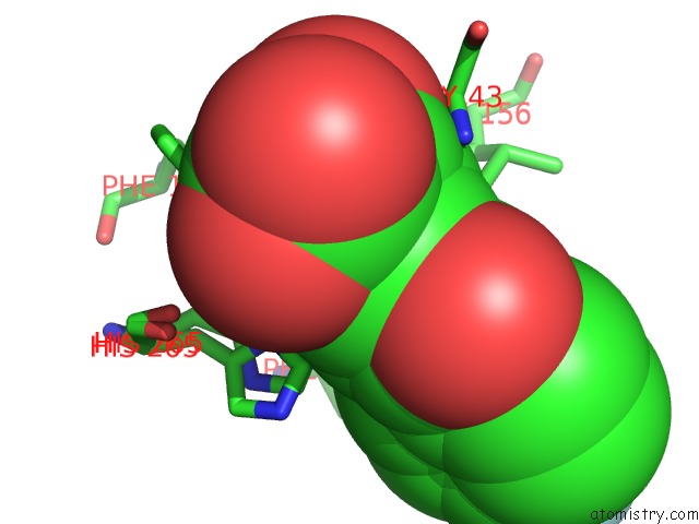
Mono view
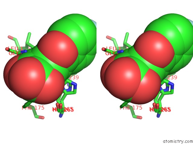
Stereo pair view
Mono view
Stereo pair view
A full contact list of Fluorine with other atoms in the F binding
site number 1 of Crystal Structure of the S112A Mutant of A C-C Hydrolase, Bphd From Burkholderia Xenovorans LB400, in Complex with 3,10-Di-Fluoro Hopda within 5.0Å range:
|
Fluorine binding site 2 out of 2 in 2rhw
Go back to
Fluorine binding site 2 out
of 2 in the Crystal Structure of the S112A Mutant of A C-C Hydrolase, Bphd From Burkholderia Xenovorans LB400, in Complex with 3,10-Di-Fluoro Hopda
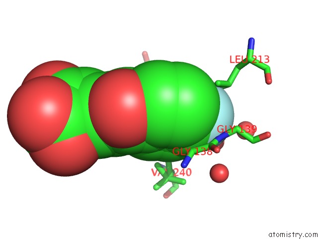
Mono view
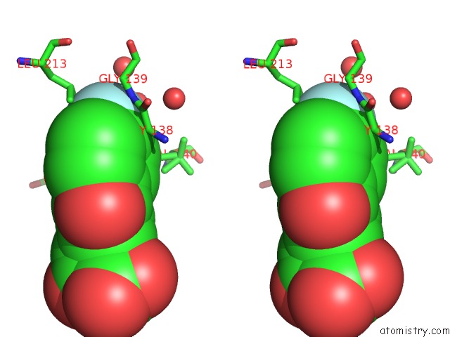
Stereo pair view
Mono view
Stereo pair view
A full contact list of Fluorine with other atoms in the F binding
site number 2 of Crystal Structure of the S112A Mutant of A C-C Hydrolase, Bphd From Burkholderia Xenovorans LB400, in Complex with 3,10-Di-Fluoro Hopda within 5.0Å range:
|
Reference:
S.Bhowmik,
G.P.Horsman,
J.T.Bolin,
L.D.Eltis.
The Molecular Basis For Inhibition of Bphd, A C-C Bond Hydrolase Involved in Polychlorinated Biphenyls Degradation: Large 3-Substituents Prevent Tautomerization. J.Biol.Chem. V. 282 36377 2007.
ISSN: ISSN 0021-9258
PubMed: 17932031
DOI: 10.1074/JBC.M707035200
Page generated: Mon Jul 14 14:19:48 2025
ISSN: ISSN 0021-9258
PubMed: 17932031
DOI: 10.1074/JBC.M707035200
Last articles
K in 3R6HK in 3Q9F
K in 3R0O
K in 3QV7
K in 3QW2
K in 3QV9
K in 3QV6
K in 3QSF
K in 3QSC
K in 3Q9C