Fluorine »
PDB 2rfn-2vfz »
2skc »
Fluorine in PDB 2skc: Pyridoxal Phosphorylase B in Complex with Fluorophosphate, Glucose and Inosine-5'-Monophosphate
Enzymatic activity of Pyridoxal Phosphorylase B in Complex with Fluorophosphate, Glucose and Inosine-5'-Monophosphate
All present enzymatic activity of Pyridoxal Phosphorylase B in Complex with Fluorophosphate, Glucose and Inosine-5'-Monophosphate:
2.4.1.1;
2.4.1.1;
Protein crystallography data
The structure of Pyridoxal Phosphorylase B in Complex with Fluorophosphate, Glucose and Inosine-5'-Monophosphate, PDB code: 2skc
was solved by
N.G.Oikonomakos,
S.E.Zographos,
K.E.Tsitsanou,
L.N.Johnson,
K.R.Acharya,
with X-Ray Crystallography technique. A brief refinement statistics is given in the table below:
| Resolution Low / High (Å) | 27.84 / 2.40 |
| Space group | P 43 21 2 |
| Cell size a, b, c (Å), α, β, γ (°) | 128.500, 128.500, 116.300, 90.00, 90.00, 90.00 |
| R / Rfree (%) | 16.7 / 21.7 |
Fluorine Binding Sites:
The binding sites of Fluorine atom in the Pyridoxal Phosphorylase B in Complex with Fluorophosphate, Glucose and Inosine-5'-Monophosphate
(pdb code 2skc). This binding sites where shown within
5.0 Angstroms radius around Fluorine atom.
In total only one binding site of Fluorine was determined in the Pyridoxal Phosphorylase B in Complex with Fluorophosphate, Glucose and Inosine-5'-Monophosphate, PDB code: 2skc:
In total only one binding site of Fluorine was determined in the Pyridoxal Phosphorylase B in Complex with Fluorophosphate, Glucose and Inosine-5'-Monophosphate, PDB code: 2skc:
Fluorine binding site 1 out of 1 in 2skc
Go back to
Fluorine binding site 1 out
of 1 in the Pyridoxal Phosphorylase B in Complex with Fluorophosphate, Glucose and Inosine-5'-Monophosphate
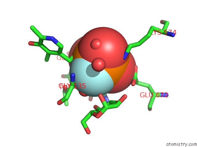
Mono view
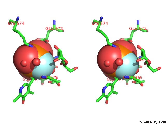
Stereo pair view
Mono view
Stereo pair view
A full contact list of Fluorine with other atoms in the F binding
site number 1 of Pyridoxal Phosphorylase B in Complex with Fluorophosphate, Glucose and Inosine-5'-Monophosphate within 5.0Å range:
|
Reference:
N.G.Oikonomakos,
S.E.Zographos,
K.E.Tsitsanou,
L.N.Johnson,
K.R.Acharya.
Activator Anion Binding Site in Pyridoxal Phosphorylase B: the Binding of Phosphite, Phosphate, and Fluorophosphate in the Crystal. Protein Sci. V. 5 2416 1996.
ISSN: ISSN 0961-8368
PubMed: 8976550
Page generated: Mon Jul 14 14:20:21 2025
ISSN: ISSN 0961-8368
PubMed: 8976550
Last articles
Mn in 9LJUMn in 9LJW
Mn in 9LJS
Mn in 9LJR
Mn in 9LJT
Mn in 9LJV
Mg in 9UA2
Mg in 9R96
Mg in 9VM1
Mg in 9P01