Fluorine »
PDB 2rfn-2vfz »
2v8y »
Fluorine in PDB 2v8y: Crystallographic and Mass Spectrometric Characterisation of EIF4E with N7-Cap Derivatives
Protein crystallography data
The structure of Crystallographic and Mass Spectrometric Characterisation of EIF4E with N7-Cap Derivatives, PDB code: 2v8y
was solved by
C.J.Brown,
I.Mcnae,
P.M.Fischer,
M.D.Walkinshaw,
with X-Ray Crystallography technique. A brief refinement statistics is given in the table below:
| Resolution Low / High (Å) | 25.30 / 2.10 |
| Space group | P 21 21 21 |
| Cell size a, b, c (Å), α, β, γ (°) | 38.438, 100.731, 135.334, 90.00, 90.00, 90.00 |
| R / Rfree (%) | 20.3 / 25 |
Fluorine Binding Sites:
The binding sites of Fluorine atom in the Crystallographic and Mass Spectrometric Characterisation of EIF4E with N7-Cap Derivatives
(pdb code 2v8y). This binding sites where shown within
5.0 Angstroms radius around Fluorine atom.
In total 2 binding sites of Fluorine where determined in the Crystallographic and Mass Spectrometric Characterisation of EIF4E with N7-Cap Derivatives, PDB code: 2v8y:
Jump to Fluorine binding site number: 1; 2;
In total 2 binding sites of Fluorine where determined in the Crystallographic and Mass Spectrometric Characterisation of EIF4E with N7-Cap Derivatives, PDB code: 2v8y:
Jump to Fluorine binding site number: 1; 2;
Fluorine binding site 1 out of 2 in 2v8y
Go back to
Fluorine binding site 1 out
of 2 in the Crystallographic and Mass Spectrometric Characterisation of EIF4E with N7-Cap Derivatives
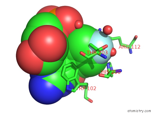
Mono view
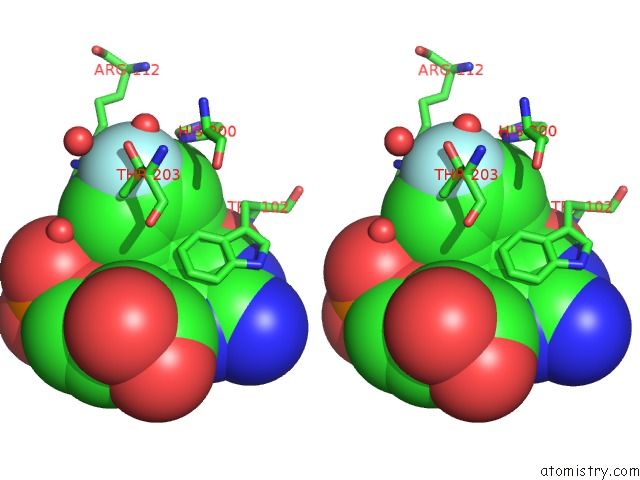
Stereo pair view
Mono view
Stereo pair view
A full contact list of Fluorine with other atoms in the F binding
site number 1 of Crystallographic and Mass Spectrometric Characterisation of EIF4E with N7-Cap Derivatives within 5.0Å range:
|
Fluorine binding site 2 out of 2 in 2v8y
Go back to
Fluorine binding site 2 out
of 2 in the Crystallographic and Mass Spectrometric Characterisation of EIF4E with N7-Cap Derivatives
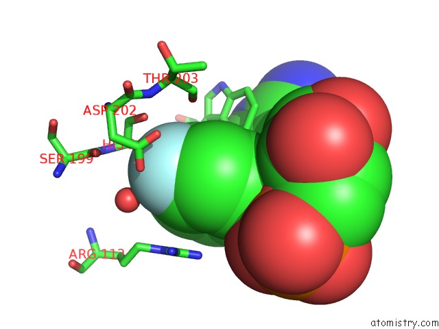
Mono view
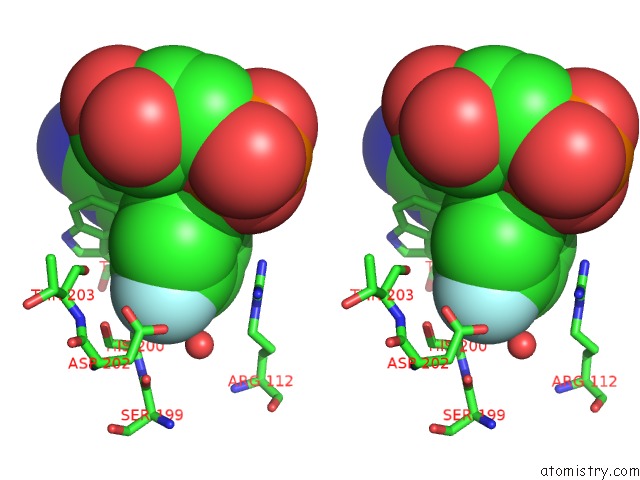
Stereo pair view
Mono view
Stereo pair view
A full contact list of Fluorine with other atoms in the F binding
site number 2 of Crystallographic and Mass Spectrometric Characterisation of EIF4E with N7-Cap Derivatives within 5.0Å range:
|
Reference:
C.J.Brown,
I.Mcnae,
P.M.Fischer,
M.D.Walkinshaw.
Crystallographic and Mass Spectrometric Characterisation of EIF4E with N(7)-Alkylated Cap Derivatives. J.Mol.Biol. V. 372 7 2007.
ISSN: ISSN 0022-2836
PubMed: 17631896
DOI: 10.1016/J.JMB.2007.06.033
Page generated: Mon Jul 14 14:23:54 2025
ISSN: ISSN 0022-2836
PubMed: 17631896
DOI: 10.1016/J.JMB.2007.06.033
Last articles
Mn in 9LJUMn in 9LJW
Mn in 9LJS
Mn in 9LJR
Mn in 9LJT
Mn in 9LJV
Mg in 9UA2
Mg in 9R96
Mg in 9VM1
Mg in 9P01