Fluorine »
PDB 2weg-2x2f »
2x1n »
Fluorine in PDB 2x1n: Truncation and Optimisation of Peptide Inhibitors of CDK2, Cyclin A Through Structure Guided Design
Enzymatic activity of Truncation and Optimisation of Peptide Inhibitors of CDK2, Cyclin A Through Structure Guided Design
All present enzymatic activity of Truncation and Optimisation of Peptide Inhibitors of CDK2, Cyclin A Through Structure Guided Design:
2.7.1.37;
2.7.1.37;
Protein crystallography data
The structure of Truncation and Optimisation of Peptide Inhibitors of CDK2, Cyclin A Through Structure Guided Design, PDB code: 2x1n
was solved by
G.Kontopidis,
M.J.Andrews,
C.Mcinnes,
A.Plater,
L.Innes,
S.Renachowski,
A.Cowan,
P.M.Fischer,
N.A.Mcintyre,
G.Griffiths,
A.L.Barnett,
A.M.Z.Slawin,
W.Jackson,
M.Thomas,
D.I.Zheleva,
S.Wang,
D.G.Blake,
N.J.Westwood,
with X-Ray Crystallography technique. A brief refinement statistics is given in the table below:
| Resolution Low / High (Å) | 30.00 / 2.75 |
| Space group | P 21 21 21 |
| Cell size a, b, c (Å), α, β, γ (°) | 74.558, 114.257, 157.277, 90.00, 90.00, 90.00 |
| R / Rfree (%) | 20 / 25.5 |
Fluorine Binding Sites:
The binding sites of Fluorine atom in the Truncation and Optimisation of Peptide Inhibitors of CDK2, Cyclin A Through Structure Guided Design
(pdb code 2x1n). This binding sites where shown within
5.0 Angstroms radius around Fluorine atom.
In total only one binding site of Fluorine was determined in the Truncation and Optimisation of Peptide Inhibitors of CDK2, Cyclin A Through Structure Guided Design, PDB code: 2x1n:
In total only one binding site of Fluorine was determined in the Truncation and Optimisation of Peptide Inhibitors of CDK2, Cyclin A Through Structure Guided Design, PDB code: 2x1n:
Fluorine binding site 1 out of 1 in 2x1n
Go back to
Fluorine binding site 1 out
of 1 in the Truncation and Optimisation of Peptide Inhibitors of CDK2, Cyclin A Through Structure Guided Design
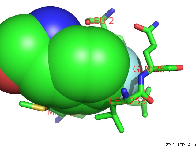
Mono view
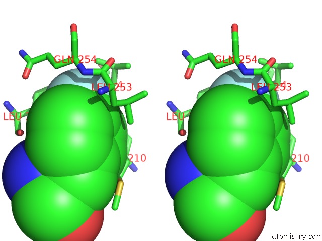
Stereo pair view
Mono view
Stereo pair view
A full contact list of Fluorine with other atoms in the F binding
site number 1 of Truncation and Optimisation of Peptide Inhibitors of CDK2, Cyclin A Through Structure Guided Design within 5.0Å range:
|
Reference:
N.A.Mcintyre,
C.Mcinnes,
G.Griffiths,
A.L.Barnett,
G.Kontopidis,
A.M.Z.Slawin,
W.Jackson,
M.Thomas,
D.I.Zheleva,
S.Wang,
D.G.Blake,
N.J.Westwood,
P.M.Fischer.
Design, Synthesis, and Evaluation of 2-Methyl- and 2-Amino-N-Aryl-4,5-Dihydrothiazolo[4,5-H]Quinazolin-8-Amines As Ring-Constrained 2-Anilino-4-(Thiazol-5-Yl)Pyrimidine Cyclin-Dependent Kinase Inhibitors. J.Med.Chem. V. 53 2136 2010.
ISSN: ISSN 0022-2623
PubMed: 20146435
DOI: 10.1021/JM901660C
Page generated: Mon Jul 14 14:45:54 2025
ISSN: ISSN 0022-2623
PubMed: 20146435
DOI: 10.1021/JM901660C
Last articles
Na in 2X7KNa in 2X6W
Na in 2X6Y
Na in 2X76
Na in 2X5X
Na in 2X2V
Na in 2X0R
Na in 2X2F
Na in 2X2E
Na in 2X1Z