Fluorine »
PDB 2x2n-2y1w »
2xvk »
Fluorine in PDB 2xvk: Crystal Structure of Alpha-Xylosidase (GH31) From Cellvibrio Japonicus in Complex with 5-Fluoro-Alpha-D-Xylopyranosyl Fluoride
Protein crystallography data
The structure of Crystal Structure of Alpha-Xylosidase (GH31) From Cellvibrio Japonicus in Complex with 5-Fluoro-Alpha-D-Xylopyranosyl Fluoride, PDB code: 2xvk
was solved by
J.Larsbrink,
A.Izumi,
F.Ibatullin,
A.Nakhai,
H.J.Gilbert,
G.J.Davies,
H.Brumer,
with X-Ray Crystallography technique. A brief refinement statistics is given in the table below:
| Resolution Low / High (Å) | 49.985 / 2.50 |
| Space group | P 63 2 2 |
| Cell size a, b, c (Å), α, β, γ (°) | 156.524, 156.524, 227.762, 90.00, 90.00, 120.00 |
| R / Rfree (%) | 19.49 / 23.97 |
Other elements in 2xvk:
The structure of Crystal Structure of Alpha-Xylosidase (GH31) From Cellvibrio Japonicus in Complex with 5-Fluoro-Alpha-D-Xylopyranosyl Fluoride also contains other interesting chemical elements:
| Chlorine | (Cl) | 4 atoms |
| Nickel | (Ni) | 1 atom |
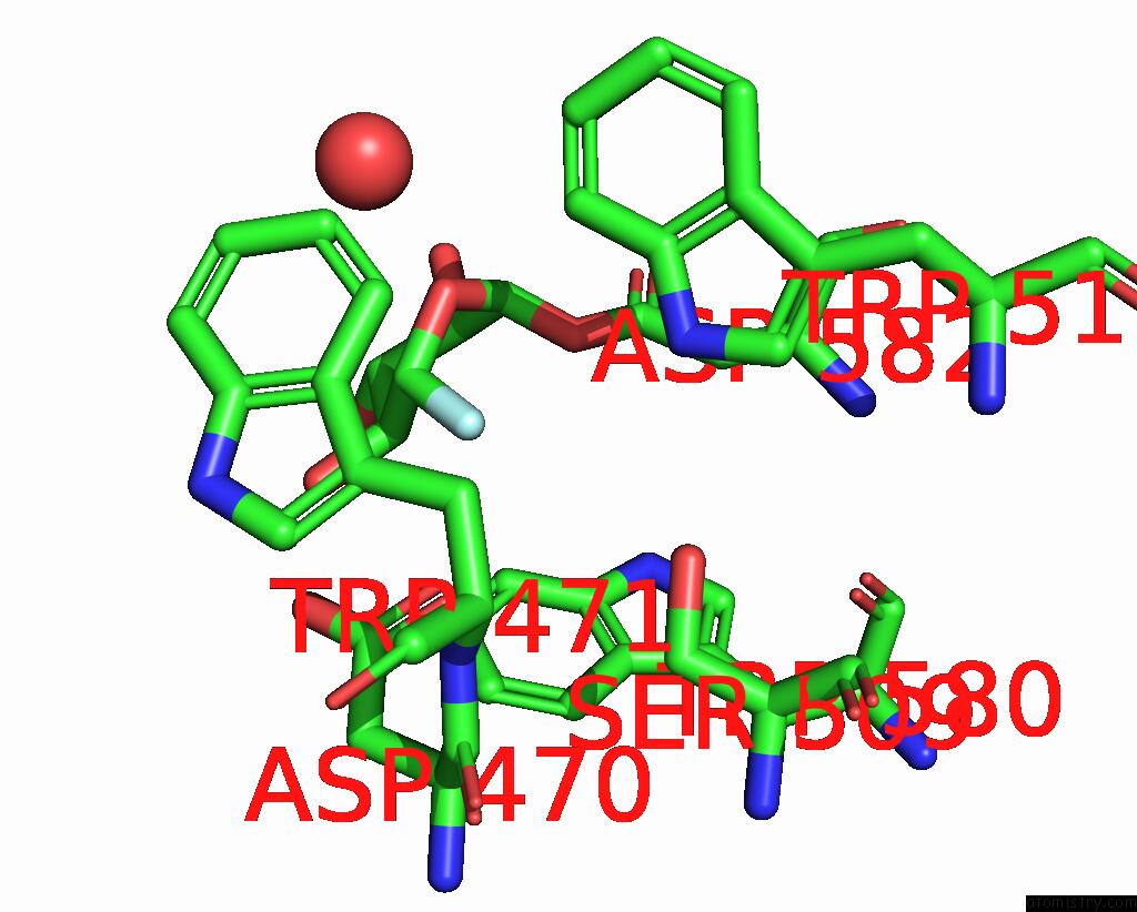
Fluorine Binding Sites:
The binding sites of Fluorine atom in the Crystal Structure of Alpha-Xylosidase (GH31) From Cellvibrio Japonicus in Complex with 5-Fluoro-Alpha-D-Xylopyranosyl Fluoride
(pdb code 2xvk). This binding sites where shown within
5.0 Angstroms radius around Fluorine atom.
In total only one binding site of Fluorine was determined in the Crystal Structure of Alpha-Xylosidase (GH31) From Cellvibrio Japonicus in Complex with 5-Fluoro-Alpha-D-Xylopyranosyl Fluoride, PDB code: 2xvk:
In total only one binding site of Fluorine was determined in the Crystal Structure of Alpha-Xylosidase (GH31) From Cellvibrio Japonicus in Complex with 5-Fluoro-Alpha-D-Xylopyranosyl Fluoride, PDB code: 2xvk:
Fluorine binding site 1 out of 1 in 2xvk
Go back to
Fluorine binding site 1 out
of 1 in the Crystal Structure of Alpha-Xylosidase (GH31) From Cellvibrio Japonicus in Complex with 5-Fluoro-Alpha-D-Xylopyranosyl Fluoride
Mono view
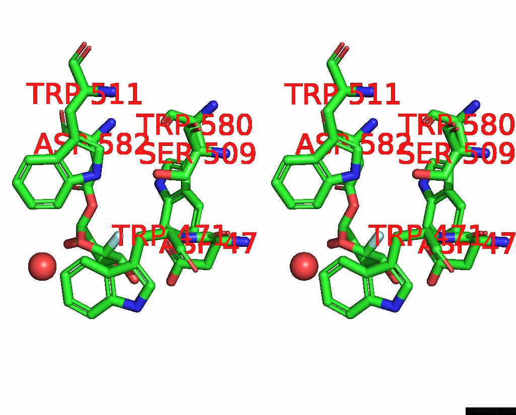
Stereo pair view
Mono view
Stereo pair view
A full contact list of Fluorine with other atoms in the F binding
site number 1 of Crystal Structure of Alpha-Xylosidase (GH31) From Cellvibrio Japonicus in Complex with 5-Fluoro-Alpha-D-Xylopyranosyl Fluoride within 5.0Å range:
|
Reference:
J.Larsbrink,
A.Izumi,
F.Ibatullin,
A.Nakhai,
H.J.Gilbert,
G.J.Davies,
H.Brumer.
Structural and Enzymatic Characterisation of A Glycoside Hydrolase Family 31 Alpha-Xylosidase From Cellvibrio Japonicus Involved in Xyloglucan Saccharification. Biochem.J. V. 436 567 2011.
ISSN: ISSN 0264-6021
PubMed: 21426303
DOI: 10.1042/BJ20110299
Page generated: Mon Jul 14 14:51:26 2025
ISSN: ISSN 0264-6021
PubMed: 21426303
DOI: 10.1042/BJ20110299
Last articles
Mg in 3CCVMg in 3CCU
Mg in 3CCS
Mg in 3CCR
Mg in 3CCQ
Mg in 3CCM
Mg in 3CCL
Mg in 3CCJ
Mg in 3CC2
Mg in 3CCE