Fluorine »
PDB 3b0q-3cct »
3ba8 »
Fluorine in PDB 3ba8: Structural Basis For the Inhibition of Bacterial Nad+ Dependent Dna Ligase
Enzymatic activity of Structural Basis For the Inhibition of Bacterial Nad+ Dependent Dna Ligase
All present enzymatic activity of Structural Basis For the Inhibition of Bacterial Nad+ Dependent Dna Ligase:
6.5.1.2;
6.5.1.2;
Protein crystallography data
The structure of Structural Basis For the Inhibition of Bacterial Nad+ Dependent Dna Ligase, PDB code: 3ba8
was solved by
C.Pinko,
with X-Ray Crystallography technique. A brief refinement statistics is given in the table below:
| Resolution Low / High (Å) | 500.00 / 1.90 |
| Space group | C 1 2 1 |
| Cell size a, b, c (Å), α, β, γ (°) | 90.496, 86.285, 56.609, 90.00, 100.94, 90.00 |
| R / Rfree (%) | 19.3 / 22.3 |
Fluorine Binding Sites:
The binding sites of Fluorine atom in the Structural Basis For the Inhibition of Bacterial Nad+ Dependent Dna Ligase
(pdb code 3ba8). This binding sites where shown within
5.0 Angstroms radius around Fluorine atom.
In total only one binding site of Fluorine was determined in the Structural Basis For the Inhibition of Bacterial Nad+ Dependent Dna Ligase, PDB code: 3ba8:
In total only one binding site of Fluorine was determined in the Structural Basis For the Inhibition of Bacterial Nad+ Dependent Dna Ligase, PDB code: 3ba8:
Fluorine binding site 1 out of 1 in 3ba8
Go back to
Fluorine binding site 1 out
of 1 in the Structural Basis For the Inhibition of Bacterial Nad+ Dependent Dna Ligase
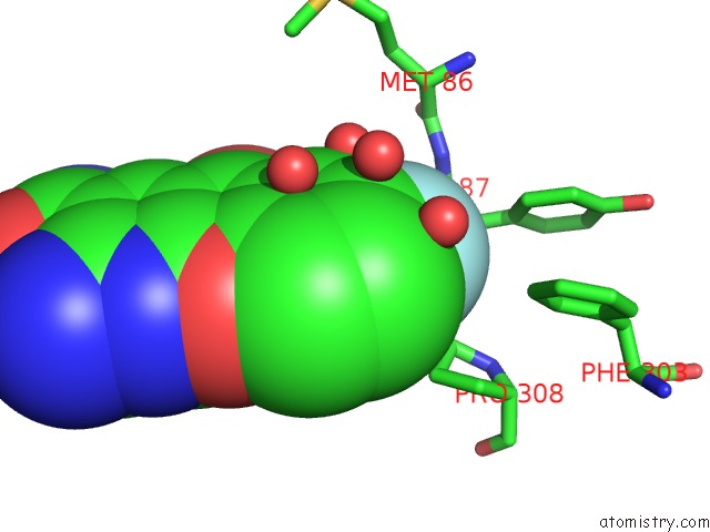
Mono view
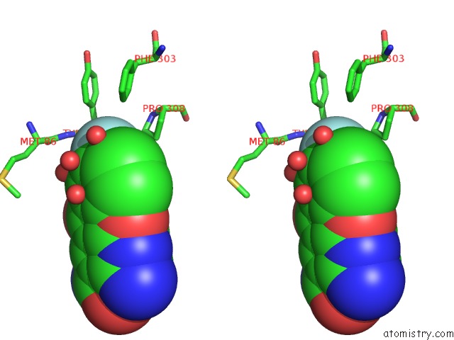
Stereo pair view
Mono view
Stereo pair view
A full contact list of Fluorine with other atoms in the F binding
site number 1 of Structural Basis For the Inhibition of Bacterial Nad+ Dependent Dna Ligase within 5.0Å range:
|
Reference:
C.Pinko,
A.Borchardt,
V.Nikulin,
Y.Su.
Structural Basis For the Inhibition of Bacterial Nad+ Dependent Dna Ligase To Be Published.
Page generated: Mon Jul 14 15:18:29 2025
Last articles
Mg in 5GAEMg in 5G0R
Mg in 5G5V
Mg in 5G3T
Mg in 5G5T
Mg in 5G5S
Mg in 5G4A
Mg in 5G57
Mg in 5G50
Mg in 5G41