Fluorine »
PDB 3fls-3g70 »
3fzr »
Fluorine in PDB 3fzr: Crystal Structure of PYK2 Complexed with Pf-431396
Enzymatic activity of Crystal Structure of PYK2 Complexed with Pf-431396
All present enzymatic activity of Crystal Structure of PYK2 Complexed with Pf-431396:
2.7.10.2;
2.7.10.2;
Protein crystallography data
The structure of Crystal Structure of PYK2 Complexed with Pf-431396, PDB code: 3fzr
was solved by
S.Han,
with X-Ray Crystallography technique. A brief refinement statistics is given in the table below:
| Resolution Low / High (Å) | 17.88 / 2.70 |
| Space group | P 4 21 2 |
| Cell size a, b, c (Å), α, β, γ (°) | 107.326, 107.326, 75.786, 90.00, 90.00, 90.00 |
| R / Rfree (%) | 21.1 / 27.4 |
Fluorine Binding Sites:
The binding sites of Fluorine atom in the Crystal Structure of PYK2 Complexed with Pf-431396
(pdb code 3fzr). This binding sites where shown within
5.0 Angstroms radius around Fluorine atom.
In total 3 binding sites of Fluorine where determined in the Crystal Structure of PYK2 Complexed with Pf-431396, PDB code: 3fzr:
Jump to Fluorine binding site number: 1; 2; 3;
In total 3 binding sites of Fluorine where determined in the Crystal Structure of PYK2 Complexed with Pf-431396, PDB code: 3fzr:
Jump to Fluorine binding site number: 1; 2; 3;
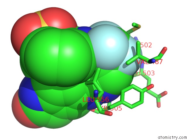
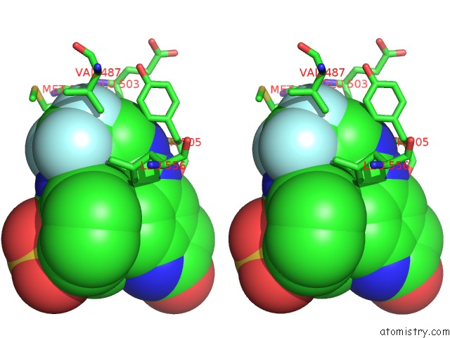
Fluorine binding site 1 out of 3 in 3fzr
Go back to
Fluorine binding site 1 out
of 3 in the Crystal Structure of PYK2 Complexed with Pf-431396
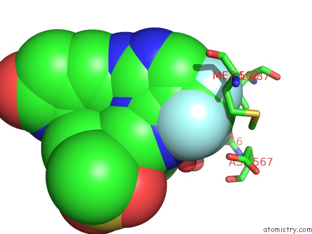
Mono view
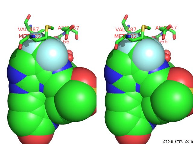
Stereo pair view
Mono view
Stereo pair view
A full contact list of Fluorine with other atoms in the F binding
site number 1 of Crystal Structure of PYK2 Complexed with Pf-431396 within 5.0Å range:
|
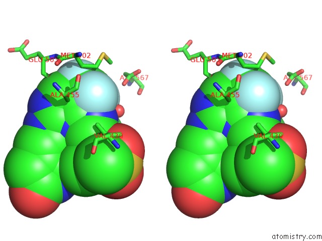
Fluorine binding site 2 out of 3 in 3fzr
Go back to
Fluorine binding site 2 out
of 3 in the Crystal Structure of PYK2 Complexed with Pf-431396
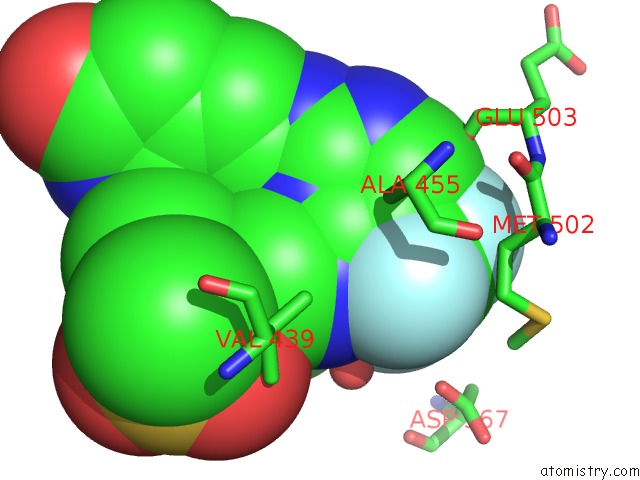
Mono view
Stereo pair view
Mono view
Stereo pair view
A full contact list of Fluorine with other atoms in the F binding
site number 2 of Crystal Structure of PYK2 Complexed with Pf-431396 within 5.0Å range:
|
Fluorine binding site 3 out of 3 in 3fzr
Go back to
Fluorine binding site 3 out
of 3 in the Crystal Structure of PYK2 Complexed with Pf-431396
Mono view
Stereo pair view
Mono view
Stereo pair view
A full contact list of Fluorine with other atoms in the F binding
site number 3 of Crystal Structure of PYK2 Complexed with Pf-431396 within 5.0Å range:
|
Reference:
S.Han,
A.Mistry,
J.S.Chang,
D.Cunningham,
M.Griffor,
P.C.Bonnette,
H.Wang,
B.A.Chrunyk,
G.E.Aspnes,
D.P.Walker,
A.D.Brosius,
L.Buckbinder.
Structural Characterization of Proline-Rich Tyrosine Kinase 2 (PYK2) Reveals A Unique (Dfg-Out) Conformation and Enables Inhibitor Design. J.Biol.Chem. V. 284 13193 2009.
ISSN: ISSN 0021-9258
PubMed: 19244237
DOI: 10.1074/JBC.M809038200
Page generated: Mon Jul 14 16:24:28 2025
ISSN: ISSN 0021-9258
PubMed: 19244237
DOI: 10.1074/JBC.M809038200
Last articles
Mn in 9LJUMn in 9LJW
Mn in 9LJS
Mn in 9LJR
Mn in 9LJT
Mn in 9LJV
Mg in 9UA2
Mg in 9R96
Mg in 9VM1
Mg in 9P01