Fluorine »
PDB 3hky-3ig6 »
3hok »
Fluorine in PDB 3hok: X-Ray Crystal Structure of Human Heme Oxygenase-1 with (2R, 4S)-2-[2-(4-Chlorophenyl)Ethyl]-2-[(1H-Imidazol-1-Yl) Methyl]-4[((5-Trifluoromethylpyridin-2-Yl)Thio)Methyl]-1,3- Dioxolane: A Novel, Inducible Binding Mode
Enzymatic activity of X-Ray Crystal Structure of Human Heme Oxygenase-1 with (2R, 4S)-2-[2-(4-Chlorophenyl)Ethyl]-2-[(1H-Imidazol-1-Yl) Methyl]-4[((5-Trifluoromethylpyridin-2-Yl)Thio)Methyl]-1,3- Dioxolane: A Novel, Inducible Binding Mode
All present enzymatic activity of X-Ray Crystal Structure of Human Heme Oxygenase-1 with (2R, 4S)-2-[2-(4-Chlorophenyl)Ethyl]-2-[(1H-Imidazol-1-Yl) Methyl]-4[((5-Trifluoromethylpyridin-2-Yl)Thio)Methyl]-1,3- Dioxolane: A Novel, Inducible Binding Mode:
1.14.99.3;
1.14.99.3;
Protein crystallography data
The structure of X-Ray Crystal Structure of Human Heme Oxygenase-1 with (2R, 4S)-2-[2-(4-Chlorophenyl)Ethyl]-2-[(1H-Imidazol-1-Yl) Methyl]-4[((5-Trifluoromethylpyridin-2-Yl)Thio)Methyl]-1,3- Dioxolane: A Novel, Inducible Binding Mode, PDB code: 3hok
was solved by
M.N.Rahman,
Z.Jia,
with X-Ray Crystallography technique. A brief refinement statistics is given in the table below:
| Resolution Low / High (Å) | 43.42 / 2.19 |
| Space group | P 1 21 1 |
| Cell size a, b, c (Å), α, β, γ (°) | 55.072, 54.592, 71.884, 90.00, 94.64, 90.00 |
| R / Rfree (%) | 22.2 / 28.9 |
Other elements in 3hok:
The structure of X-Ray Crystal Structure of Human Heme Oxygenase-1 with (2R, 4S)-2-[2-(4-Chlorophenyl)Ethyl]-2-[(1H-Imidazol-1-Yl) Methyl]-4[((5-Trifluoromethylpyridin-2-Yl)Thio)Methyl]-1,3- Dioxolane: A Novel, Inducible Binding Mode also contains other interesting chemical elements:
| Iron | (Fe) | 2 atoms |
| Chlorine | (Cl) | 1 atom |
Fluorine Binding Sites:
The binding sites of Fluorine atom in the X-Ray Crystal Structure of Human Heme Oxygenase-1 with (2R, 4S)-2-[2-(4-Chlorophenyl)Ethyl]-2-[(1H-Imidazol-1-Yl) Methyl]-4[((5-Trifluoromethylpyridin-2-Yl)Thio)Methyl]-1,3- Dioxolane: A Novel, Inducible Binding Mode
(pdb code 3hok). This binding sites where shown within
5.0 Angstroms radius around Fluorine atom.
In total 3 binding sites of Fluorine where determined in the X-Ray Crystal Structure of Human Heme Oxygenase-1 with (2R, 4S)-2-[2-(4-Chlorophenyl)Ethyl]-2-[(1H-Imidazol-1-Yl) Methyl]-4[((5-Trifluoromethylpyridin-2-Yl)Thio)Methyl]-1,3- Dioxolane: A Novel, Inducible Binding Mode, PDB code: 3hok:
Jump to Fluorine binding site number: 1; 2; 3;
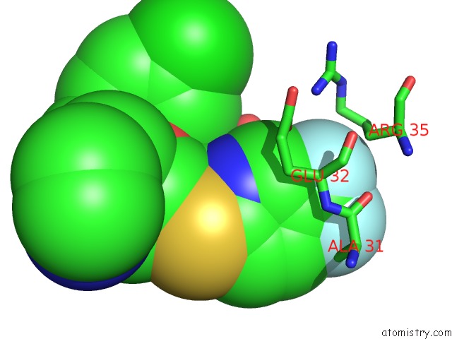
Fluorine binding site 1 out of 3 in 3hok
Go back to
Fluorine binding site 1 out
of 3 in the X-Ray Crystal Structure of Human Heme Oxygenase-1 with (2R, 4S)-2-[2-(4-Chlorophenyl)Ethyl]-2-[(1H-Imidazol-1-Yl) Methyl]-4[((5-Trifluoromethylpyridin-2-Yl)Thio)Methyl]-1,3- Dioxolane: A Novel, Inducible Binding Mode
Mono view

Stereo pair view
Mono view
Stereo pair view
A full contact list of Fluorine with other atoms in the F binding
site number 1 of X-Ray Crystal Structure of Human Heme Oxygenase-1 with (2R, 4S)-2-[2-(4-Chlorophenyl)Ethyl]-2-[(1H-Imidazol-1-Yl) Methyl]-4[((5-Trifluoromethylpyridin-2-Yl)Thio)Methyl]-1,3- Dioxolane: A Novel, Inducible Binding Mode within 5.0Å range:
|
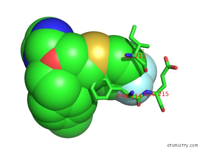
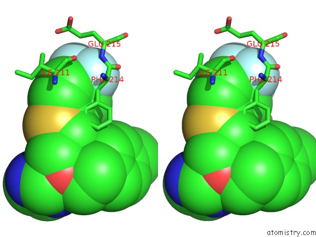
Fluorine binding site 2 out of 3 in 3hok
Go back to
Fluorine binding site 2 out
of 3 in the X-Ray Crystal Structure of Human Heme Oxygenase-1 with (2R, 4S)-2-[2-(4-Chlorophenyl)Ethyl]-2-[(1H-Imidazol-1-Yl) Methyl]-4[((5-Trifluoromethylpyridin-2-Yl)Thio)Methyl]-1,3- Dioxolane: A Novel, Inducible Binding Mode
Mono view
Stereo pair view
Mono view
Stereo pair view
A full contact list of Fluorine with other atoms in the F binding
site number 2 of X-Ray Crystal Structure of Human Heme Oxygenase-1 with (2R, 4S)-2-[2-(4-Chlorophenyl)Ethyl]-2-[(1H-Imidazol-1-Yl) Methyl]-4[((5-Trifluoromethylpyridin-2-Yl)Thio)Methyl]-1,3- Dioxolane: A Novel, Inducible Binding Mode within 5.0Å range:
|
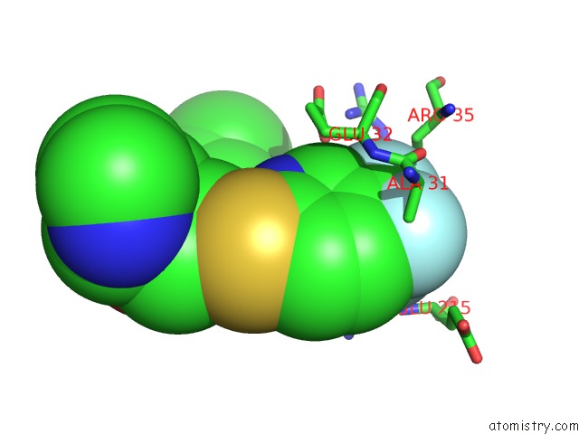
Fluorine binding site 3 out of 3 in 3hok
Go back to
Fluorine binding site 3 out
of 3 in the X-Ray Crystal Structure of Human Heme Oxygenase-1 with (2R, 4S)-2-[2-(4-Chlorophenyl)Ethyl]-2-[(1H-Imidazol-1-Yl) Methyl]-4[((5-Trifluoromethylpyridin-2-Yl)Thio)Methyl]-1,3- Dioxolane: A Novel, Inducible Binding Mode
Mono view

Stereo pair view
Mono view
Stereo pair view
A full contact list of Fluorine with other atoms in the F binding
site number 3 of X-Ray Crystal Structure of Human Heme Oxygenase-1 with (2R, 4S)-2-[2-(4-Chlorophenyl)Ethyl]-2-[(1H-Imidazol-1-Yl) Methyl]-4[((5-Trifluoromethylpyridin-2-Yl)Thio)Methyl]-1,3- Dioxolane: A Novel, Inducible Binding Mode within 5.0Å range:
|
Reference:
M.N.Rahman,
J.Z.Vlahakis,
D.Vukomanovic,
W.A.Szarek,
K.Nakatsu,
Z.Jia.
X-Ray Crystal Structure of Human Heme Oxygenase-1 with (2R,4S)-2-[2-(4-Chlorophenyl)Ethyl]-2-[(1H- Imidazol-1-Yl)Methyl]-4[((5-Trifluoromethylpyridin- 2-Yl)Thio)Methyl]-1,3-Dioxolane: A Novel, Inducible Binding Mode. J.Med.Chem. V. 52 4946 2009.
ISSN: ISSN 0022-2623
PubMed: 19601578
DOI: 10.1021/JM900434F
Page generated: Mon Jul 14 16:54:40 2025
ISSN: ISSN 0022-2623
PubMed: 19601578
DOI: 10.1021/JM900434F
Last articles
Na in 3CCONa in 3CC2
Na in 3CCE
Na in 3CC7
Na in 3CC4
Na in 3CC9
Na in 3CBC
Na in 3CBT
Na in 3C9F
Na in 3CB8