Fluorine »
PDB 3kqn-3l8s »
3ktu »
Fluorine in PDB 3ktu: Structure of Human 8-Oxoguanine Glycosylase 1 Bound to Fluorninated Oxog-Containing Dna
Enzymatic activity of Structure of Human 8-Oxoguanine Glycosylase 1 Bound to Fluorninated Oxog-Containing Dna
All present enzymatic activity of Structure of Human 8-Oxoguanine Glycosylase 1 Bound to Fluorninated Oxog-Containing Dna:
4.2.99.18;
4.2.99.18;
Protein crystallography data
The structure of Structure of Human 8-Oxoguanine Glycosylase 1 Bound to Fluorninated Oxog-Containing Dna, PDB code: 3ktu
was solved by
G.L.Verdine,
S.M.Lee,
with X-Ray Crystallography technique. A brief refinement statistics is given in the table below:
| Resolution Low / High (Å) | 25.21 / 2.30 |
| Space group | P 65 2 2 |
| Cell size a, b, c (Å), α, β, γ (°) | 92.400, 92.400, 212.500, 90.00, 90.00, 120.00 |
| R / Rfree (%) | 22.5 / 25.7 |
Other elements in 3ktu:
The structure of Structure of Human 8-Oxoguanine Glycosylase 1 Bound to Fluorninated Oxog-Containing Dna also contains other interesting chemical elements:
| Calcium | (Ca) | 2 atoms |
Fluorine Binding Sites:
The binding sites of Fluorine atom in the Structure of Human 8-Oxoguanine Glycosylase 1 Bound to Fluorninated Oxog-Containing Dna
(pdb code 3ktu). This binding sites where shown within
5.0 Angstroms radius around Fluorine atom.
In total only one binding site of Fluorine was determined in the Structure of Human 8-Oxoguanine Glycosylase 1 Bound to Fluorninated Oxog-Containing Dna, PDB code: 3ktu:
In total only one binding site of Fluorine was determined in the Structure of Human 8-Oxoguanine Glycosylase 1 Bound to Fluorninated Oxog-Containing Dna, PDB code: 3ktu:
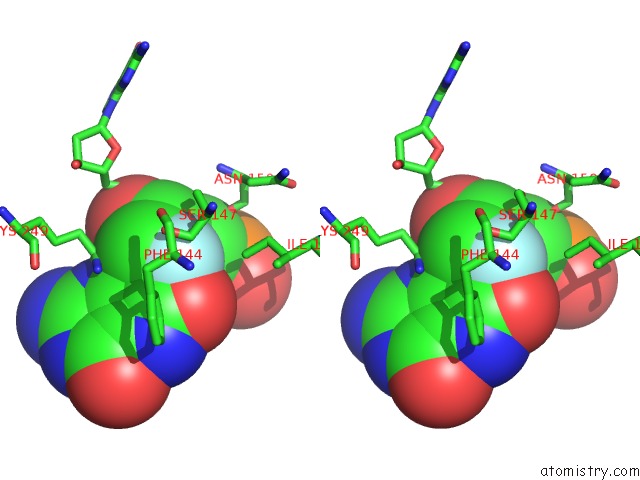
Fluorine binding site 1 out of 1 in 3ktu
Go back to
Fluorine binding site 1 out
of 1 in the Structure of Human 8-Oxoguanine Glycosylase 1 Bound to Fluorninated Oxog-Containing Dna

Mono view
Stereo pair view
Mono view
Stereo pair view
A full contact list of Fluorine with other atoms in the F binding
site number 1 of Structure of Human 8-Oxoguanine Glycosylase 1 Bound to Fluorninated Oxog-Containing Dna within 5.0Å range:
|
Reference:
G.L.Verdine,
S.M.Lee.
Structural Investigation of HOGG1 Bound to A Fluorinated Oxog Analog To Be Published.
Page generated: Mon Jul 14 17:34:28 2025
Last articles
Mg in 3OLPMg in 3OLX
Mg in 3OLV
Mg in 3OJU
Mg in 3OKS
Mg in 3OJS
Mg in 3OIV
Mg in 3OIW
Mg in 3OI7
Mg in 3OIU