Fluorine »
PDB 3l8v-3lxp »
3len »
Fluorine in PDB 3len: Human Aldose Reductase Mutant T113S Complexed with Zopolrestat
Enzymatic activity of Human Aldose Reductase Mutant T113S Complexed with Zopolrestat
All present enzymatic activity of Human Aldose Reductase Mutant T113S Complexed with Zopolrestat:
1.1.1.21;
1.1.1.21;
Protein crystallography data
The structure of Human Aldose Reductase Mutant T113S Complexed with Zopolrestat, PDB code: 3len
was solved by
C.Koch,
A.Heine,
G.Klebe,
with X-Ray Crystallography technique. A brief refinement statistics is given in the table below:
| Resolution Low / High (Å) | 10.00 / 1.21 |
| Space group | P 1 |
| Cell size a, b, c (Å), α, β, γ (°) | 40.000, 47.070, 47.410, 75.81, 67.58, 77.32 |
| R / Rfree (%) | 12.3 / 16.1 |
Fluorine Binding Sites:
The binding sites of Fluorine atom in the Human Aldose Reductase Mutant T113S Complexed with Zopolrestat
(pdb code 3len). This binding sites where shown within
5.0 Angstroms radius around Fluorine atom.
In total 3 binding sites of Fluorine where determined in the Human Aldose Reductase Mutant T113S Complexed with Zopolrestat, PDB code: 3len:
Jump to Fluorine binding site number: 1; 2; 3;
In total 3 binding sites of Fluorine where determined in the Human Aldose Reductase Mutant T113S Complexed with Zopolrestat, PDB code: 3len:
Jump to Fluorine binding site number: 1; 2; 3;
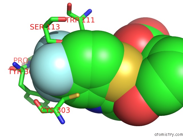
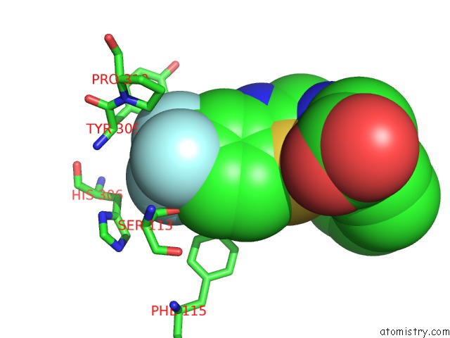
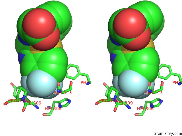
Fluorine binding site 1 out of 3 in 3len
Go back to
Fluorine binding site 1 out
of 3 in the Human Aldose Reductase Mutant T113S Complexed with Zopolrestat
Mono view
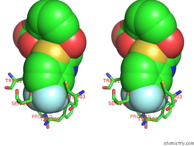
Stereo pair view
Mono view
Stereo pair view
A full contact list of Fluorine with other atoms in the F binding
site number 1 of Human Aldose Reductase Mutant T113S Complexed with Zopolrestat within 5.0Å range:
|
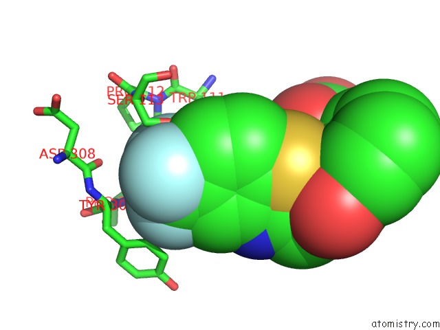
Fluorine binding site 2 out of 3 in 3len
Go back to
Fluorine binding site 2 out
of 3 in the Human Aldose Reductase Mutant T113S Complexed with Zopolrestat
Mono view
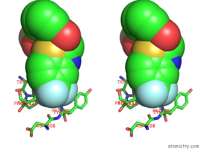
Stereo pair view
Mono view
Stereo pair view
A full contact list of Fluorine with other atoms in the F binding
site number 2 of Human Aldose Reductase Mutant T113S Complexed with Zopolrestat within 5.0Å range:
|
Fluorine binding site 3 out of 3 in 3len
Go back to
Fluorine binding site 3 out
of 3 in the Human Aldose Reductase Mutant T113S Complexed with Zopolrestat
Mono view
Stereo pair view
Mono view
Stereo pair view
A full contact list of Fluorine with other atoms in the F binding
site number 3 of Human Aldose Reductase Mutant T113S Complexed with Zopolrestat within 5.0Å range:
|
Reference:
C.Koch,
A.Heine,
G.Klebe.
Ligand-Induced Fit Affects Binding Modes and Provokes Changes in Crystal Packing of Aldose Reductase Biochim.Biophys.Acta V.1810 879 2011.
ISSN: ISSN 0006-3002
PubMed: 21684320
DOI: 10.1016/J.BBAGEN.2011.06.001
Page generated: Mon Jul 14 17:44:51 2025
ISSN: ISSN 0006-3002
PubMed: 21684320
DOI: 10.1016/J.BBAGEN.2011.06.001
Last articles
K in 7KURK in 7KUQ
K in 7KLU
K in 7KLP
K in 7KNC
K in 7KLV
K in 7KMP
K in 7KDY
K in 7KLB
K in 7KD9