Fluorine »
PDB 3lz3-3n0n »
3mba »
Fluorine in PDB 3mba: Aplysia Limacina Myoglobin. Crystallographic Analysis at 1.6 Angstroms Resolution
Protein crystallography data
The structure of Aplysia Limacina Myoglobin. Crystallographic Analysis at 1.6 Angstroms Resolution, PDB code: 3mba
was solved by
M.Bolognesi,
S.Onesti,
G.Gatti,
A.Coda,
P.Ascenzi,
M.Brunori,
with X-Ray Crystallography technique. A brief refinement statistics is given in the table below:
| Resolution Low / High (Å) | N/A / 2.00 |
| Space group | P 21 21 21 |
| Cell size a, b, c (Å), α, β, γ (°) | 52.980, 70.700, 32.500, 90.00, 90.00, 90.00 |
| R / Rfree (%) | n/a / n/a |
Other elements in 3mba:
The structure of Aplysia Limacina Myoglobin. Crystallographic Analysis at 1.6 Angstroms Resolution also contains other interesting chemical elements:
| Iron | (Fe) | 1 atom |
Fluorine Binding Sites:
The binding sites of Fluorine atom in the Aplysia Limacina Myoglobin. Crystallographic Analysis at 1.6 Angstroms Resolution
(pdb code 3mba). This binding sites where shown within
5.0 Angstroms radius around Fluorine atom.
In total only one binding site of Fluorine was determined in the Aplysia Limacina Myoglobin. Crystallographic Analysis at 1.6 Angstroms Resolution, PDB code: 3mba:
In total only one binding site of Fluorine was determined in the Aplysia Limacina Myoglobin. Crystallographic Analysis at 1.6 Angstroms Resolution, PDB code: 3mba:
Fluorine binding site 1 out of 1 in 3mba
Go back to
Fluorine binding site 1 out
of 1 in the Aplysia Limacina Myoglobin. Crystallographic Analysis at 1.6 Angstroms Resolution
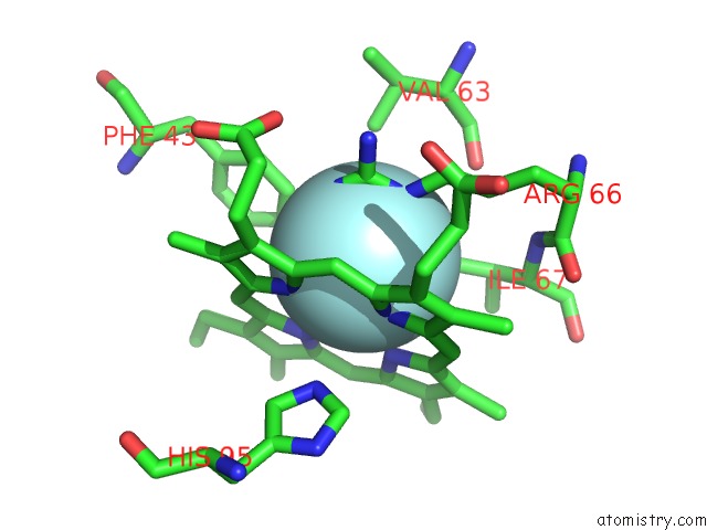
Mono view
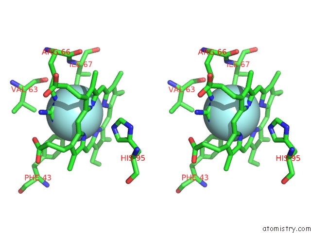
Stereo pair view
Mono view
Stereo pair view
A full contact list of Fluorine with other atoms in the F binding
site number 1 of Aplysia Limacina Myoglobin. Crystallographic Analysis at 1.6 Angstroms Resolution within 5.0Å range:
|
Reference:
M.Bolognesi,
S.Onesti,
G.Gatti,
A.Coda,
P.Ascenzi,
M.Brunori.
Aplysia Limacina Myoglobin. Crystallographic Analysis at 1.6 A Resolution. J.Mol.Biol. V. 205 529 1989.
ISSN: ISSN 0022-2836
PubMed: 2926816
DOI: 10.1016/0022-2836(89)90224-6
Page generated: Wed Jul 31 20:40:51 2024
ISSN: ISSN 0022-2836
PubMed: 2926816
DOI: 10.1016/0022-2836(89)90224-6
Last articles
Zn in 9MJ5Zn in 9HNW
Zn in 9G0L
Zn in 9FNE
Zn in 9DZN
Zn in 9E0I
Zn in 9D32
Zn in 9DAK
Zn in 8ZXC
Zn in 8ZUF