Fluorine »
PDB 3nlz-3og7 »
3odv »
Fluorine in PDB 3odv: X-Ray Structure of Kaliotoxin By Racemic Protein Crystallography
Protein crystallography data
The structure of X-Ray Structure of Kaliotoxin By Racemic Protein Crystallography, PDB code: 3odv
was solved by
B.L.Pentelute,
K.Mandal,
Z.P.Gates,
M.R.Sawaya,
T.O.Yeates,
S.B.H.Kent,
with X-Ray Crystallography technique. A brief refinement statistics is given in the table below:
| Resolution Low / High (Å) | 38.13 / 0.95 |
| Space group | P -1 |
| Cell size a, b, c (Å), α, β, γ (°) | 25.150, 30.511, 41.097, 109.39, 97.39, 97.09 |
| R / Rfree (%) | 18.8 / 20.3 |
Fluorine Binding Sites:
The binding sites of Fluorine atom in the X-Ray Structure of Kaliotoxin By Racemic Protein Crystallography
(pdb code 3odv). This binding sites where shown within
5.0 Angstroms radius around Fluorine atom.
In total 6 binding sites of Fluorine where determined in the X-Ray Structure of Kaliotoxin By Racemic Protein Crystallography, PDB code: 3odv:
Jump to Fluorine binding site number: 1; 2; 3; 4; 5; 6;
In total 6 binding sites of Fluorine where determined in the X-Ray Structure of Kaliotoxin By Racemic Protein Crystallography, PDB code: 3odv:
Jump to Fluorine binding site number: 1; 2; 3; 4; 5; 6;
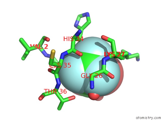
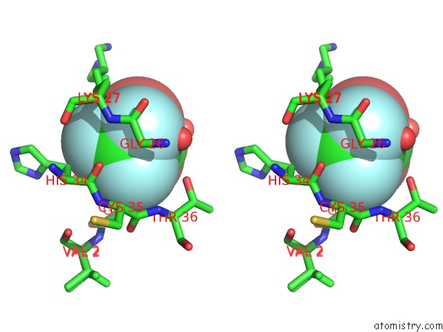
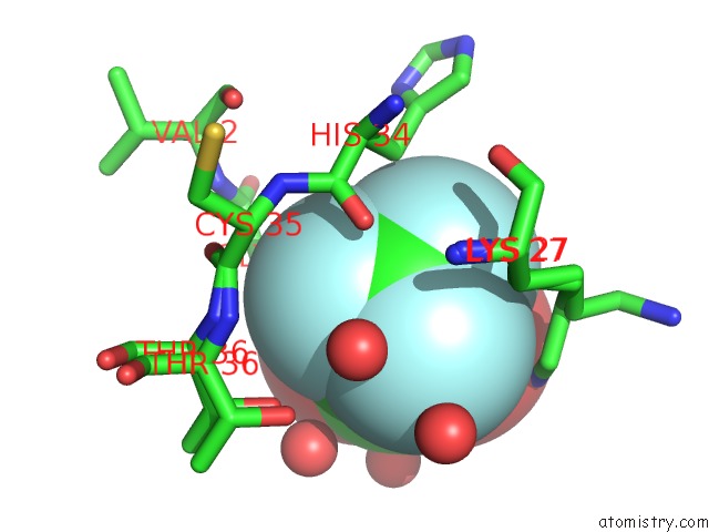
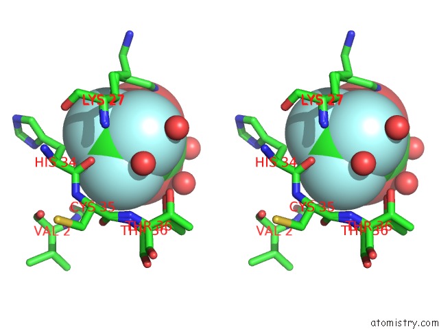
Fluorine binding site 1 out of 6 in 3odv
Go back to
Fluorine binding site 1 out
of 6 in the X-Ray Structure of Kaliotoxin By Racemic Protein Crystallography
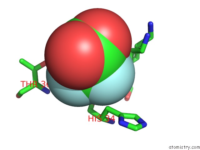
Mono view
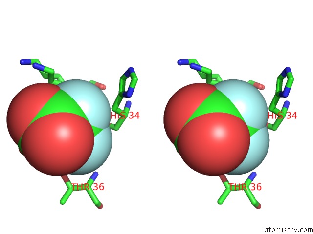
Stereo pair view
Mono view
Stereo pair view
A full contact list of Fluorine with other atoms in the F binding
site number 1 of X-Ray Structure of Kaliotoxin By Racemic Protein Crystallography within 5.0Å range:
|
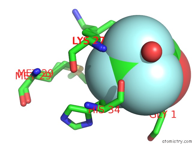
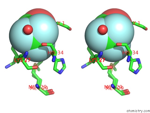
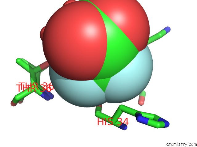
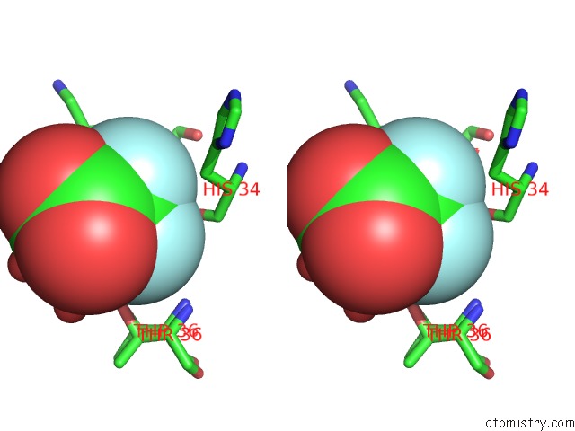
Fluorine binding site 2 out of 6 in 3odv
Go back to
Fluorine binding site 2 out
of 6 in the X-Ray Structure of Kaliotoxin By Racemic Protein Crystallography
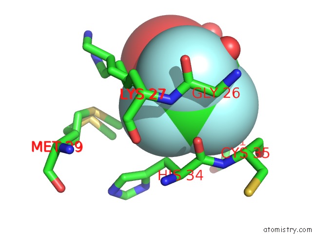
Mono view
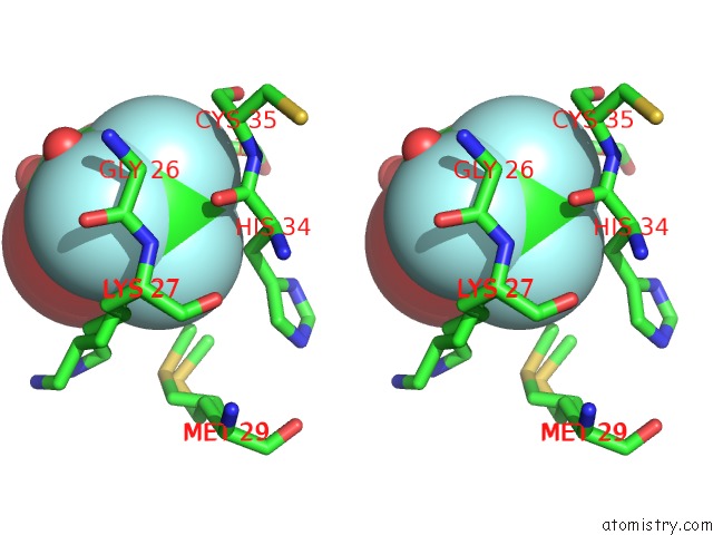
Stereo pair view
Mono view
Stereo pair view
A full contact list of Fluorine with other atoms in the F binding
site number 2 of X-Ray Structure of Kaliotoxin By Racemic Protein Crystallography within 5.0Å range:
|
Fluorine binding site 3 out of 6 in 3odv
Go back to
Fluorine binding site 3 out
of 6 in the X-Ray Structure of Kaliotoxin By Racemic Protein Crystallography
Mono view
Stereo pair view
Mono view
Stereo pair view
A full contact list of Fluorine with other atoms in the F binding
site number 3 of X-Ray Structure of Kaliotoxin By Racemic Protein Crystallography within 5.0Å range:
|
Fluorine binding site 4 out of 6 in 3odv
Go back to
Fluorine binding site 4 out
of 6 in the X-Ray Structure of Kaliotoxin By Racemic Protein Crystallography
Mono view
Stereo pair view
Mono view
Stereo pair view
A full contact list of Fluorine with other atoms in the F binding
site number 4 of X-Ray Structure of Kaliotoxin By Racemic Protein Crystallography within 5.0Å range:
|
Fluorine binding site 5 out of 6 in 3odv
Go back to
Fluorine binding site 5 out
of 6 in the X-Ray Structure of Kaliotoxin By Racemic Protein Crystallography
Mono view
Stereo pair view
Mono view
Stereo pair view
A full contact list of Fluorine with other atoms in the F binding
site number 5 of X-Ray Structure of Kaliotoxin By Racemic Protein Crystallography within 5.0Å range:
|
Fluorine binding site 6 out of 6 in 3odv
Go back to
Fluorine binding site 6 out
of 6 in the X-Ray Structure of Kaliotoxin By Racemic Protein Crystallography
Mono view
Stereo pair view
Mono view
Stereo pair view
A full contact list of Fluorine with other atoms in the F binding
site number 6 of X-Ray Structure of Kaliotoxin By Racemic Protein Crystallography within 5.0Å range:
|
Reference:
B.L.Pentelute,
K.Mandal,
Z.P.Gates,
M.R.Sawaya,
T.O.Yeates,
S.B.Kent.
Total Chemical Synthesis and X-Ray Structure of Kaliotoxin By Racemic Protein Crystallography. Chem.Commun.(Camb.) V. 46 8174 2010.
ISSN: ISSN 1359-7345
PubMed: 20877851
DOI: 10.1039/C0CC03148H
Page generated: Mon Jul 14 18:17:55 2025
ISSN: ISSN 1359-7345
PubMed: 20877851
DOI: 10.1039/C0CC03148H
Last articles
Mn in 9LJUMn in 9LJW
Mn in 9LJS
Mn in 9LJR
Mn in 9LJT
Mn in 9LJV
Mg in 9UA2
Mg in 9R96
Mg in 9VM1
Mg in 9P01