Fluorine »
PDB 3ptm-3qri »
3qi3 »
Fluorine in PDB 3qi3: Crystal Structure of PDE9A(Q453E) in Complex with Inhibitor BAY73-6691
Enzymatic activity of Crystal Structure of PDE9A(Q453E) in Complex with Inhibitor BAY73-6691
All present enzymatic activity of Crystal Structure of PDE9A(Q453E) in Complex with Inhibitor BAY73-6691:
3.1.4.35;
3.1.4.35;
Protein crystallography data
The structure of Crystal Structure of PDE9A(Q453E) in Complex with Inhibitor BAY73-6691, PDB code: 3qi3
was solved by
J.Hou,
J.Xu,
M.Liu,
R.Zhao,
H.Lou,
H.Ke,
with X-Ray Crystallography technique. A brief refinement statistics is given in the table below:
| Resolution Low / High (Å) | 30.00 / 2.30 |
| Space group | P 41 21 2 |
| Cell size a, b, c (Å), α, β, γ (°) | 103.127, 103.127, 270.441, 90.00, 90.00, 90.00 |
| R / Rfree (%) | 22.2 / 24.2 |
Other elements in 3qi3:
The structure of Crystal Structure of PDE9A(Q453E) in Complex with Inhibitor BAY73-6691 also contains other interesting chemical elements:
| Magnesium | (Mg) | 2 atoms |
| Zinc | (Zn) | 2 atoms |
| Chlorine | (Cl) | 2 atoms |
Fluorine Binding Sites:
The binding sites of Fluorine atom in the Crystal Structure of PDE9A(Q453E) in Complex with Inhibitor BAY73-6691
(pdb code 3qi3). This binding sites where shown within
5.0 Angstroms radius around Fluorine atom.
In total 6 binding sites of Fluorine where determined in the Crystal Structure of PDE9A(Q453E) in Complex with Inhibitor BAY73-6691, PDB code: 3qi3:
Jump to Fluorine binding site number: 1; 2; 3; 4; 5; 6;
In total 6 binding sites of Fluorine where determined in the Crystal Structure of PDE9A(Q453E) in Complex with Inhibitor BAY73-6691, PDB code: 3qi3:
Jump to Fluorine binding site number: 1; 2; 3; 4; 5; 6;
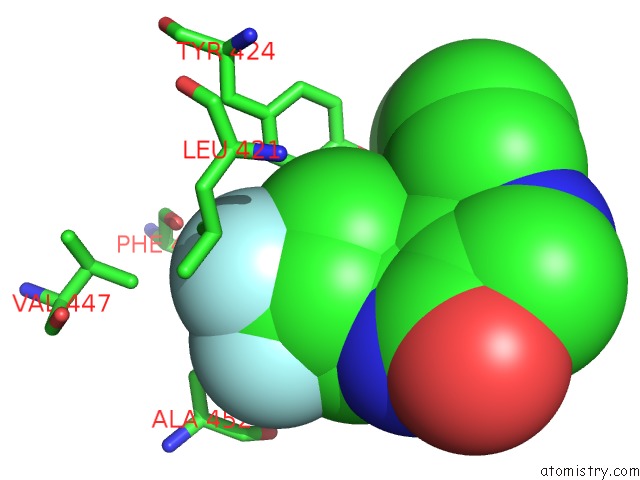
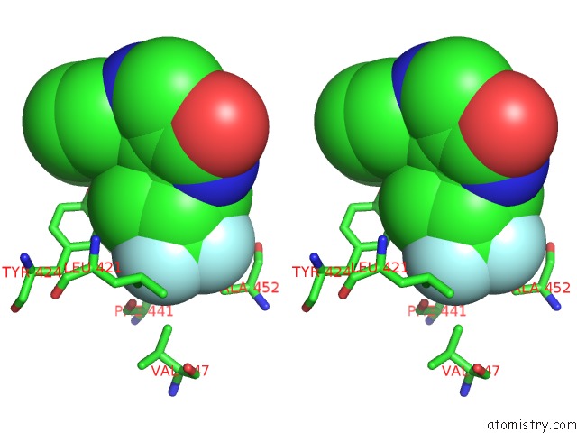
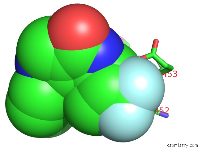
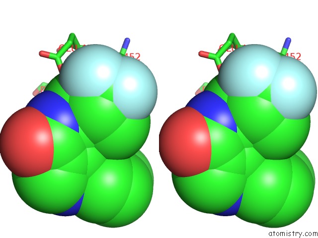
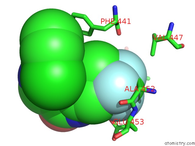
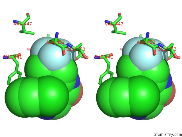
Fluorine binding site 1 out of 6 in 3qi3
Go back to
Fluorine binding site 1 out
of 6 in the Crystal Structure of PDE9A(Q453E) in Complex with Inhibitor BAY73-6691
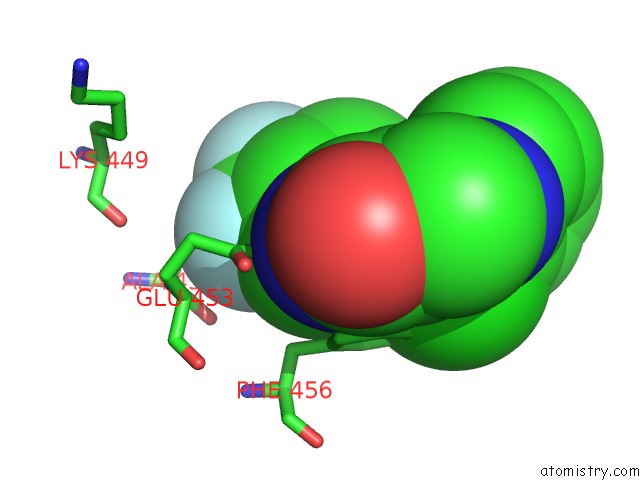
Mono view
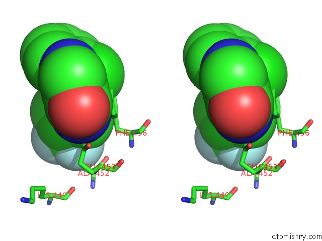
Stereo pair view
Mono view
Stereo pair view
A full contact list of Fluorine with other atoms in the F binding
site number 1 of Crystal Structure of PDE9A(Q453E) in Complex with Inhibitor BAY73-6691 within 5.0Å range:
|
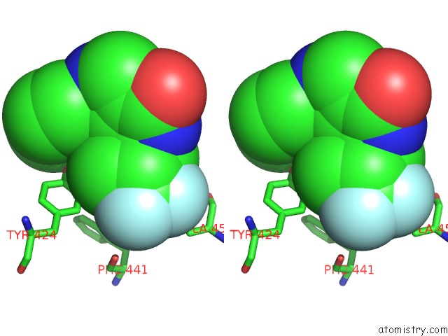
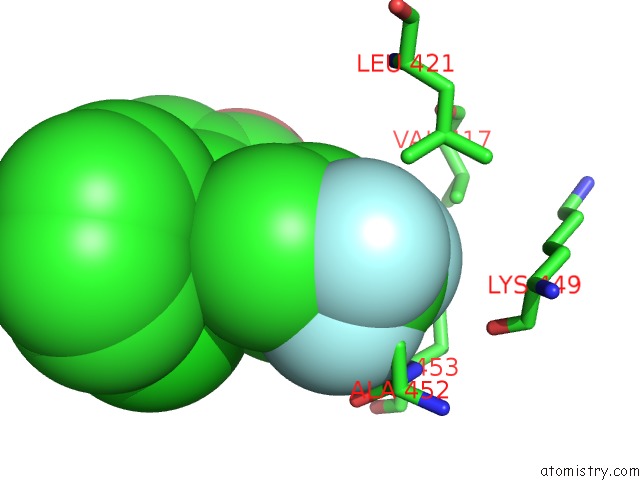
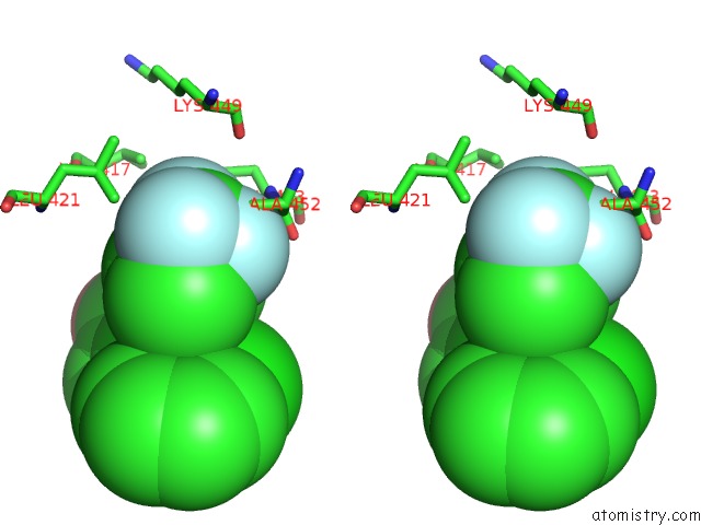
Fluorine binding site 2 out of 6 in 3qi3
Go back to
Fluorine binding site 2 out
of 6 in the Crystal Structure of PDE9A(Q453E) in Complex with Inhibitor BAY73-6691
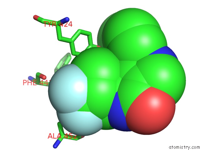
Mono view
Stereo pair view
Mono view
Stereo pair view
A full contact list of Fluorine with other atoms in the F binding
site number 2 of Crystal Structure of PDE9A(Q453E) in Complex with Inhibitor BAY73-6691 within 5.0Å range:
|
Fluorine binding site 3 out of 6 in 3qi3
Go back to
Fluorine binding site 3 out
of 6 in the Crystal Structure of PDE9A(Q453E) in Complex with Inhibitor BAY73-6691
Mono view
Stereo pair view
Mono view
Stereo pair view
A full contact list of Fluorine with other atoms in the F binding
site number 3 of Crystal Structure of PDE9A(Q453E) in Complex with Inhibitor BAY73-6691 within 5.0Å range:
|
Fluorine binding site 4 out of 6 in 3qi3
Go back to
Fluorine binding site 4 out
of 6 in the Crystal Structure of PDE9A(Q453E) in Complex with Inhibitor BAY73-6691
Mono view
Stereo pair view
Mono view
Stereo pair view
A full contact list of Fluorine with other atoms in the F binding
site number 4 of Crystal Structure of PDE9A(Q453E) in Complex with Inhibitor BAY73-6691 within 5.0Å range:
|
Fluorine binding site 5 out of 6 in 3qi3
Go back to
Fluorine binding site 5 out
of 6 in the Crystal Structure of PDE9A(Q453E) in Complex with Inhibitor BAY73-6691
Mono view
Stereo pair view
Mono view
Stereo pair view
A full contact list of Fluorine with other atoms in the F binding
site number 5 of Crystal Structure of PDE9A(Q453E) in Complex with Inhibitor BAY73-6691 within 5.0Å range:
|
Fluorine binding site 6 out of 6 in 3qi3
Go back to
Fluorine binding site 6 out
of 6 in the Crystal Structure of PDE9A(Q453E) in Complex with Inhibitor BAY73-6691
Mono view
Stereo pair view
Mono view
Stereo pair view
A full contact list of Fluorine with other atoms in the F binding
site number 6 of Crystal Structure of PDE9A(Q453E) in Complex with Inhibitor BAY73-6691 within 5.0Å range:
|
Reference:
J.Hou,
J.Xu,
M.Liu,
R.Zhao,
H.B.Luo,
H.Ke.
Structural Asymmetry of Phosphodiesterase-9, Potential Protonation of A Glutamic Acid, and Role of the Invariant Glutamine. Plos One V. 6 18092 2011.
ISSN: ESSN 1932-6203
PubMed: 21483814
DOI: 10.1371/JOURNAL.PONE.0018092
Page generated: Mon Jul 14 18:57:53 2025
ISSN: ESSN 1932-6203
PubMed: 21483814
DOI: 10.1371/JOURNAL.PONE.0018092
Last articles
Mg in 6KCCMg in 6KCA
Mg in 6KC0
Mg in 6KBZ
Mg in 6KAL
Mg in 6KAK
Mg in 6KBD
Mg in 6K9V
Mg in 6K83
Mg in 6K8K