Fluorine »
PDB 3rjc-3s9y »
3rx2 »
Fluorine in PDB 3rx2: Crystal Structure of Human Aldose Reductase Complexed with Sulindac Sulfone
Enzymatic activity of Crystal Structure of Human Aldose Reductase Complexed with Sulindac Sulfone
All present enzymatic activity of Crystal Structure of Human Aldose Reductase Complexed with Sulindac Sulfone:
1.1.1.21;
1.1.1.21;
Protein crystallography data
The structure of Crystal Structure of Human Aldose Reductase Complexed with Sulindac Sulfone, PDB code: 3rx2
was solved by
X.Zheng,
J.Chen,
H.Luo,
X.Hu,
with X-Ray Crystallography technique. A brief refinement statistics is given in the table below:
| Resolution Low / High (Å) | 24.11 / 1.90 |
| Space group | P 1 21 1 |
| Cell size a, b, c (Å), α, β, γ (°) | 47.187, 67.081, 49.357, 90.00, 91.94, 90.00 |
| R / Rfree (%) | 14.1 / 19.4 |
Fluorine Binding Sites:
The binding sites of Fluorine atom in the Crystal Structure of Human Aldose Reductase Complexed with Sulindac Sulfone
(pdb code 3rx2). This binding sites where shown within
5.0 Angstroms radius around Fluorine atom.
In total only one binding site of Fluorine was determined in the Crystal Structure of Human Aldose Reductase Complexed with Sulindac Sulfone, PDB code: 3rx2:
In total only one binding site of Fluorine was determined in the Crystal Structure of Human Aldose Reductase Complexed with Sulindac Sulfone, PDB code: 3rx2:
Fluorine binding site 1 out of 1 in 3rx2
Go back to
Fluorine binding site 1 out
of 1 in the Crystal Structure of Human Aldose Reductase Complexed with Sulindac Sulfone
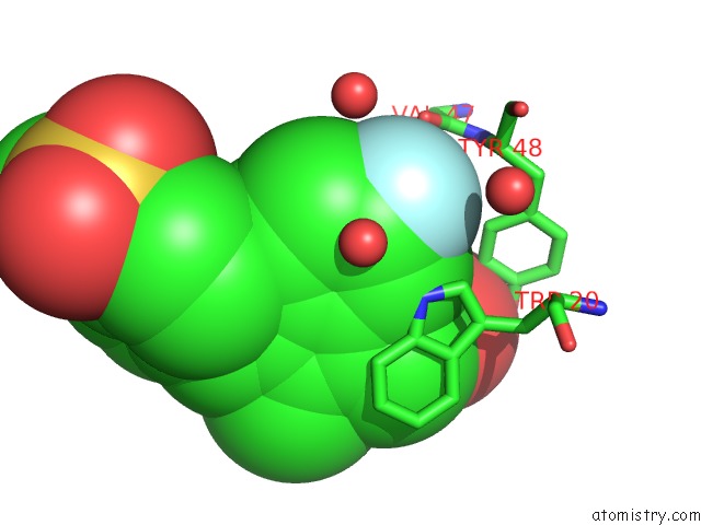
Mono view
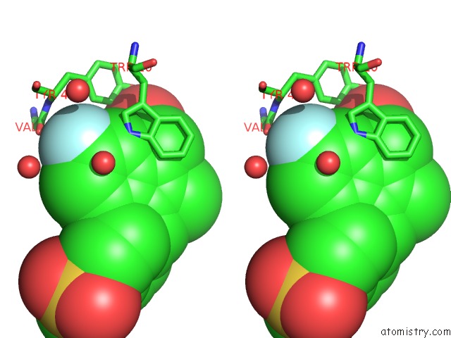
Stereo pair view
Mono view
Stereo pair view
A full contact list of Fluorine with other atoms in the F binding
site number 1 of Crystal Structure of Human Aldose Reductase Complexed with Sulindac Sulfone within 5.0Å range:
|
Reference:
X.Zheng,
L.Zhang,
J.Zhai,
Y.Chen,
H.Luo,
X.Hu.
The Molecular Basis For Inhibition of Sulindac and Its Metabolites Towards Human Aldose Reductase Febs Lett. V. 586 55 2012.
ISSN: ISSN 0014-5793
PubMed: 22155003
DOI: 10.1016/J.FEBSLET.2011.11.023
Page generated: Mon Jul 14 19:13:52 2025
ISSN: ISSN 0014-5793
PubMed: 22155003
DOI: 10.1016/J.FEBSLET.2011.11.023
Last articles
Na in 1A61Na in 1A5S
Na in 1A5G
Na in 1A4W
Na in 1A46
Na in 1A50
Na in 194L
Na in 1A2C
Na in 191D
Na in 1A3D