Fluorine »
PDB 3sb0-3sym »
3spk »
Fluorine in PDB 3spk: Tipranavir in Complex with A Human Immunodeficiency Virus Type 1 Protease Variant
Enzymatic activity of Tipranavir in Complex with A Human Immunodeficiency Virus Type 1 Protease Variant
All present enzymatic activity of Tipranavir in Complex with A Human Immunodeficiency Virus Type 1 Protease Variant:
3.4.23.16;
3.4.23.16;
Protein crystallography data
The structure of Tipranavir in Complex with A Human Immunodeficiency Virus Type 1 Protease Variant, PDB code: 3spk
was solved by
Y.Wang,
Z.Liu,
J.S.Brunzelle,
I.A.Kovari,
L.C.Kovari,
with X-Ray Crystallography technique. A brief refinement statistics is given in the table below:
| Resolution Low / High (Å) | 29.52 / 1.24 |
| Space group | P 61 |
| Cell size a, b, c (Å), α, β, γ (°) | 63.119, 63.119, 83.540, 90.00, 90.00, 120.00 |
| R / Rfree (%) | 17.4 / 22.7 |
Fluorine Binding Sites:
The binding sites of Fluorine atom in the Tipranavir in Complex with A Human Immunodeficiency Virus Type 1 Protease Variant
(pdb code 3spk). This binding sites where shown within
5.0 Angstroms radius around Fluorine atom.
In total 6 binding sites of Fluorine where determined in the Tipranavir in Complex with A Human Immunodeficiency Virus Type 1 Protease Variant, PDB code: 3spk:
Jump to Fluorine binding site number: 1; 2; 3; 4; 5; 6;
In total 6 binding sites of Fluorine where determined in the Tipranavir in Complex with A Human Immunodeficiency Virus Type 1 Protease Variant, PDB code: 3spk:
Jump to Fluorine binding site number: 1; 2; 3; 4; 5; 6;
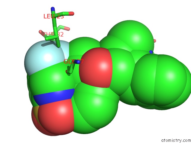
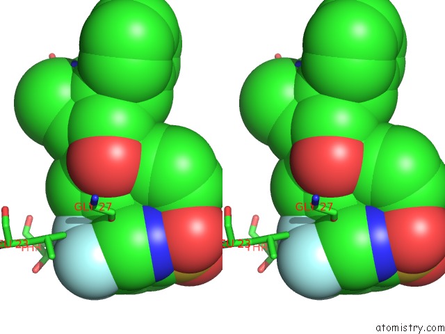
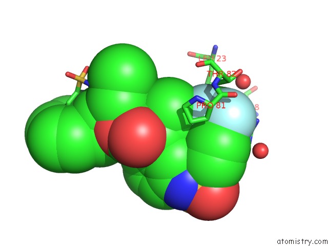
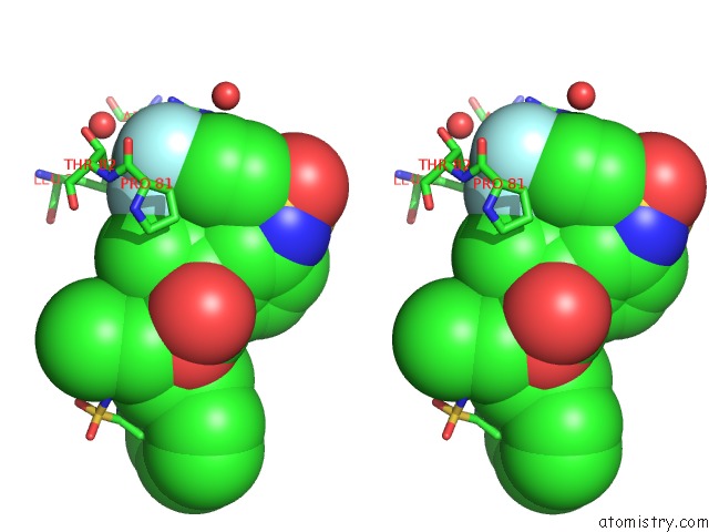
Fluorine binding site 1 out of 6 in 3spk
Go back to
Fluorine binding site 1 out
of 6 in the Tipranavir in Complex with A Human Immunodeficiency Virus Type 1 Protease Variant
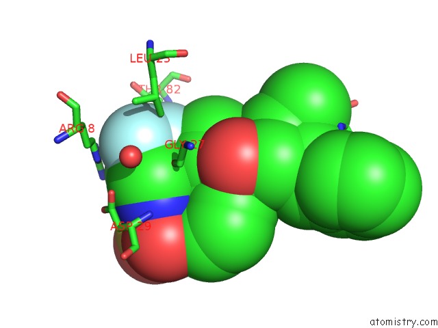
Mono view
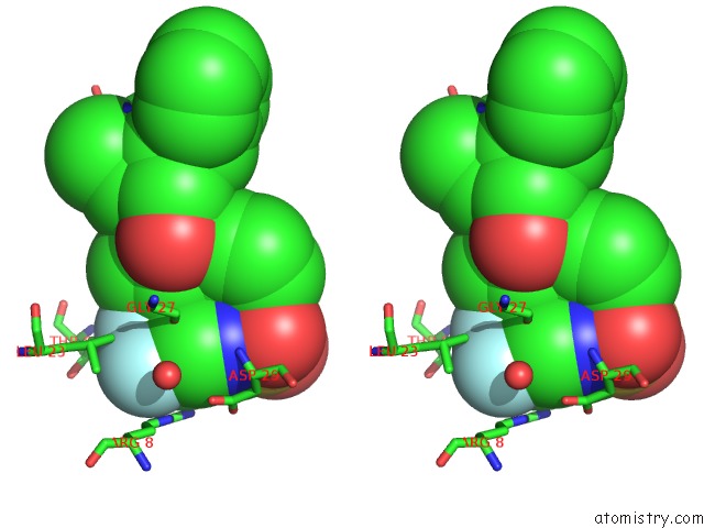
Stereo pair view
Mono view
Stereo pair view
A full contact list of Fluorine with other atoms in the F binding
site number 1 of Tipranavir in Complex with A Human Immunodeficiency Virus Type 1 Protease Variant within 5.0Å range:
|
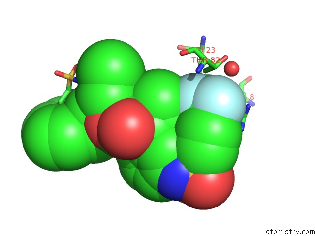
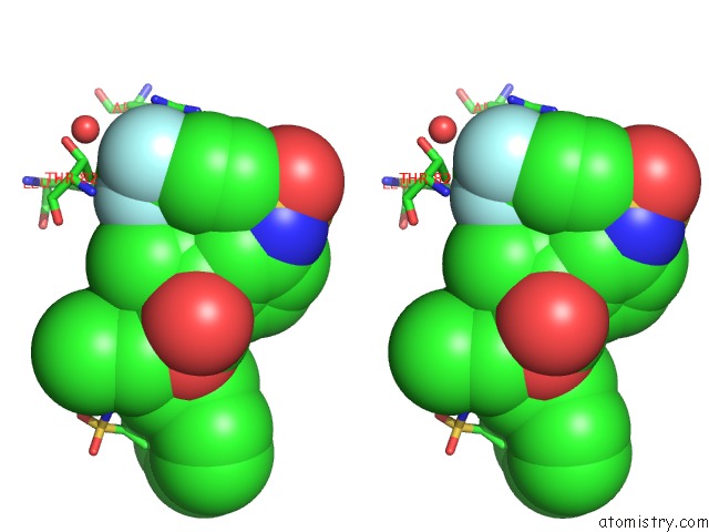
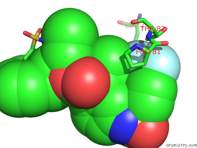
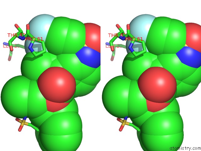
Fluorine binding site 2 out of 6 in 3spk
Go back to
Fluorine binding site 2 out
of 6 in the Tipranavir in Complex with A Human Immunodeficiency Virus Type 1 Protease Variant
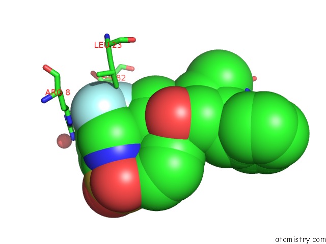
Mono view
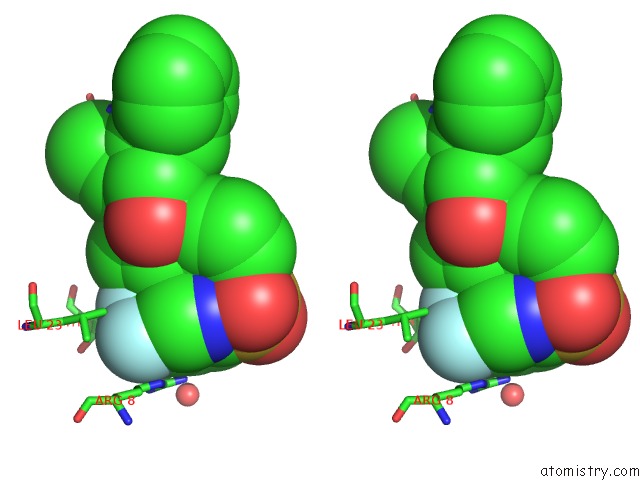
Stereo pair view
Mono view
Stereo pair view
A full contact list of Fluorine with other atoms in the F binding
site number 2 of Tipranavir in Complex with A Human Immunodeficiency Virus Type 1 Protease Variant within 5.0Å range:
|
Fluorine binding site 3 out of 6 in 3spk
Go back to
Fluorine binding site 3 out
of 6 in the Tipranavir in Complex with A Human Immunodeficiency Virus Type 1 Protease Variant
Mono view
Stereo pair view
Mono view
Stereo pair view
A full contact list of Fluorine with other atoms in the F binding
site number 3 of Tipranavir in Complex with A Human Immunodeficiency Virus Type 1 Protease Variant within 5.0Å range:
|
Fluorine binding site 4 out of 6 in 3spk
Go back to
Fluorine binding site 4 out
of 6 in the Tipranavir in Complex with A Human Immunodeficiency Virus Type 1 Protease Variant
Mono view
Stereo pair view
Mono view
Stereo pair view
A full contact list of Fluorine with other atoms in the F binding
site number 4 of Tipranavir in Complex with A Human Immunodeficiency Virus Type 1 Protease Variant within 5.0Å range:
|
Fluorine binding site 5 out of 6 in 3spk
Go back to
Fluorine binding site 5 out
of 6 in the Tipranavir in Complex with A Human Immunodeficiency Virus Type 1 Protease Variant
Mono view
Stereo pair view
Mono view
Stereo pair view
A full contact list of Fluorine with other atoms in the F binding
site number 5 of Tipranavir in Complex with A Human Immunodeficiency Virus Type 1 Protease Variant within 5.0Å range:
|
Fluorine binding site 6 out of 6 in 3spk
Go back to
Fluorine binding site 6 out
of 6 in the Tipranavir in Complex with A Human Immunodeficiency Virus Type 1 Protease Variant
Mono view
Stereo pair view
Mono view
Stereo pair view
A full contact list of Fluorine with other atoms in the F binding
site number 6 of Tipranavir in Complex with A Human Immunodeficiency Virus Type 1 Protease Variant within 5.0Å range:
|
Reference:
Y.Wang,
Z.Liu,
J.S.Brunzelle,
I.A.Kovari,
T.G.Dewdney,
S.J.Reiter,
L.C.Kovari.
The Higher Barrier of Darunavir and Tipranavir Resistance For Hiv-1 Protease. Biochem.Biophys.Res.Commun. V. 412 737 2011.
ISSN: ISSN 0006-291X
PubMed: 21871444
DOI: 10.1016/J.BBRC.2011.08.045
Page generated: Mon Jul 14 19:21:52 2025
ISSN: ISSN 0006-291X
PubMed: 21871444
DOI: 10.1016/J.BBRC.2011.08.045
Last articles
K in 9FYEK in 9FT7
K in 9FQ1
K in 9FM9
K in 9EX3
K in 9F90
K in 9ES6
K in 9EWD
K in 9ETN
K in 9ESI