Fluorine »
PDB 3sb0-3sym »
3sym »
Fluorine in PDB 3sym: Glycogen Phosphorylase B in Complex with 3 -C-(Hydroxymethyl)-Beta-D- Glucopyranonucleoside of 5-Fluorouracil
Enzymatic activity of Glycogen Phosphorylase B in Complex with 3 -C-(Hydroxymethyl)-Beta-D- Glucopyranonucleoside of 5-Fluorouracil
All present enzymatic activity of Glycogen Phosphorylase B in Complex with 3 -C-(Hydroxymethyl)-Beta-D- Glucopyranonucleoside of 5-Fluorouracil:
2.4.1.1;
2.4.1.1;
Protein crystallography data
The structure of Glycogen Phosphorylase B in Complex with 3 -C-(Hydroxymethyl)-Beta-D- Glucopyranonucleoside of 5-Fluorouracil, PDB code: 3sym
was solved by
V.T.Skamnaki,
A.L.Katsandi,
M.Kontou,
D.D.Leonidas,
with X-Ray Crystallography technique. A brief refinement statistics is given in the table below:
| Resolution Low / High (Å) | 13.86 / 2.40 |
| Space group | P 43 21 2 |
| Cell size a, b, c (Å), α, β, γ (°) | 128.324, 128.324, 116.461, 90.00, 90.00, 90.00 |
| R / Rfree (%) | 16 / 20.3 |
Fluorine Binding Sites:
The binding sites of Fluorine atom in the Glycogen Phosphorylase B in Complex with 3 -C-(Hydroxymethyl)-Beta-D- Glucopyranonucleoside of 5-Fluorouracil
(pdb code 3sym). This binding sites where shown within
5.0 Angstroms radius around Fluorine atom.
In total only one binding site of Fluorine was determined in the Glycogen Phosphorylase B in Complex with 3 -C-(Hydroxymethyl)-Beta-D- Glucopyranonucleoside of 5-Fluorouracil, PDB code: 3sym:
In total only one binding site of Fluorine was determined in the Glycogen Phosphorylase B in Complex with 3 -C-(Hydroxymethyl)-Beta-D- Glucopyranonucleoside of 5-Fluorouracil, PDB code: 3sym:
Fluorine binding site 1 out of 1 in 3sym
Go back to
Fluorine binding site 1 out
of 1 in the Glycogen Phosphorylase B in Complex with 3 -C-(Hydroxymethyl)-Beta-D- Glucopyranonucleoside of 5-Fluorouracil
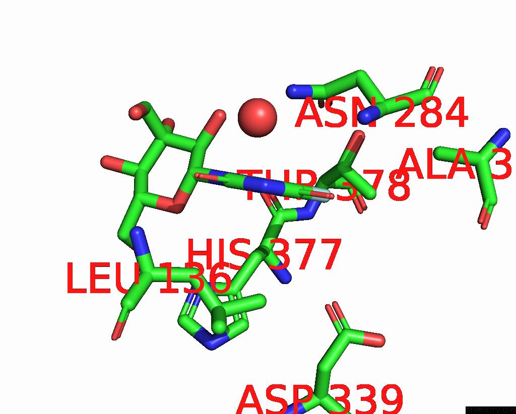
Mono view
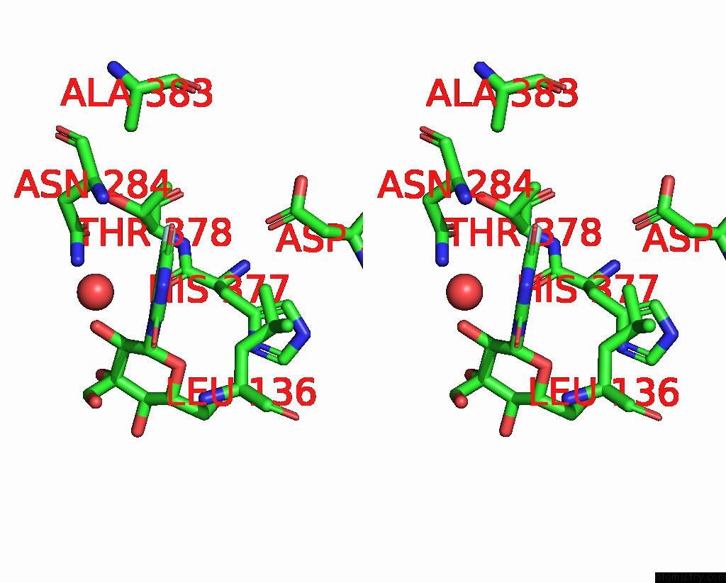
Stereo pair view
Mono view
Stereo pair view
A full contact list of Fluorine with other atoms in the F binding
site number 1 of Glycogen Phosphorylase B in Complex with 3 -C-(Hydroxymethyl)-Beta-D- Glucopyranonucleoside of 5-Fluorouracil within 5.0Å range:
|
Reference:
S.Manta,
A.Xipnitou,
C.Kiritsis,
A.L.Kantsadi,
J.M.Hayes,
V.T.Skamnaki,
C.Lamprakis,
M.Kontou,
P.Zoumpoulakis,
S.E.Zographos,
D.D.Leonidas,
D.Komiotis.
3'-Axial Ch(2) Oh Substitution on Glucopyranose Does Not Increase Glycogen Phosphorylase Inhibitory Potency. Qm/Mm-Pbsa Calculations Suggest Why. Chem.Biol.Drug Des. V. 79 663 2012.
ISSN: ISSN 1747-0277
PubMed: 22296957
DOI: 10.1111/J.1747-0285.2012.01349.X
Page generated: Wed Jul 31 22:39:10 2024
ISSN: ISSN 1747-0277
PubMed: 22296957
DOI: 10.1111/J.1747-0285.2012.01349.X
Last articles
Br in 4PBSBr in 4PA4
Br in 4PA9
Br in 4PBR
Br in 4P9P
Br in 4PA3
Br in 4P9O
Br in 4P6J
Br in 4P6K
Br in 4P4G