Fluorine »
PDB 3syn-3u2o »
3tu1 »
Fluorine in PDB 3tu1: Exhaustive Fluorine Scanning Towards Potent P53-MDM2 Antagonist
Protein crystallography data
The structure of Exhaustive Fluorine Scanning Towards Potent P53-MDM2 Antagonist, PDB code: 3tu1
was solved by
S.Wolf,
Y.Huang,
D.Koes,
G.M.Popowicz,
C.J.Camacho,
T.A.Holak,
A.Doemling,
with X-Ray Crystallography technique. A brief refinement statistics is given in the table below:
| Resolution Low / High (Å) | 20.00 / 1.60 |
| Space group | I 2 2 2 |
| Cell size a, b, c (Å), α, β, γ (°) | 49.950, 59.250, 82.950, 90.00, 90.00, 90.00 |
| R / Rfree (%) | 20.5 / 24.5 |
Other elements in 3tu1:
The structure of Exhaustive Fluorine Scanning Towards Potent P53-MDM2 Antagonist also contains other interesting chemical elements:
| Chlorine | (Cl) | 1 atom |
Fluorine Binding Sites:
The binding sites of Fluorine atom in the Exhaustive Fluorine Scanning Towards Potent P53-MDM2 Antagonist
(pdb code 3tu1). This binding sites where shown within
5.0 Angstroms radius around Fluorine atom.
In total 2 binding sites of Fluorine where determined in the Exhaustive Fluorine Scanning Towards Potent P53-MDM2 Antagonist, PDB code: 3tu1:
Jump to Fluorine binding site number: 1; 2;
In total 2 binding sites of Fluorine where determined in the Exhaustive Fluorine Scanning Towards Potent P53-MDM2 Antagonist, PDB code: 3tu1:
Jump to Fluorine binding site number: 1; 2;
Fluorine binding site 1 out of 2 in 3tu1
Go back to
Fluorine binding site 1 out
of 2 in the Exhaustive Fluorine Scanning Towards Potent P53-MDM2 Antagonist
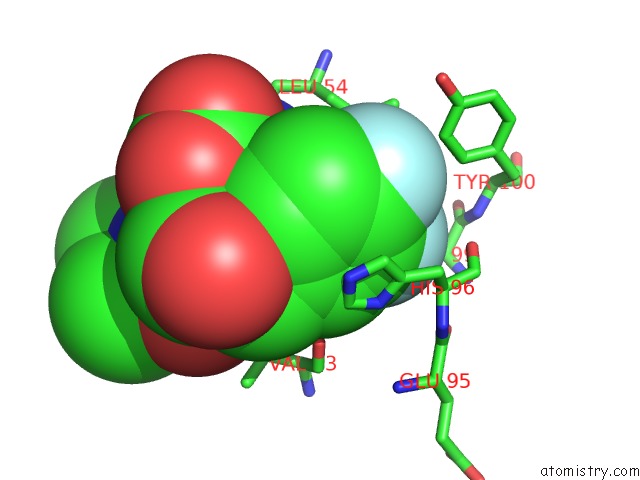
Mono view
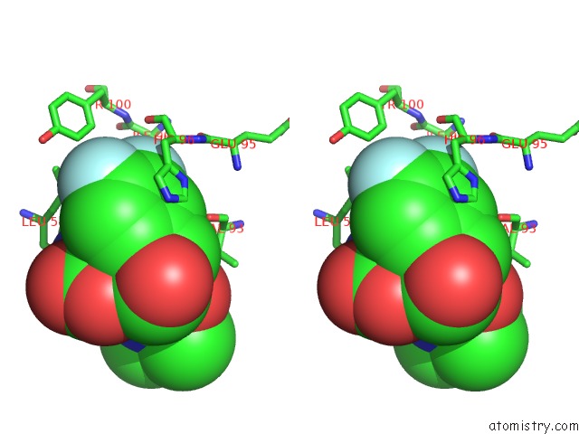
Stereo pair view
Mono view
Stereo pair view
A full contact list of Fluorine with other atoms in the F binding
site number 1 of Exhaustive Fluorine Scanning Towards Potent P53-MDM2 Antagonist within 5.0Å range:
|
Fluorine binding site 2 out of 2 in 3tu1
Go back to
Fluorine binding site 2 out
of 2 in the Exhaustive Fluorine Scanning Towards Potent P53-MDM2 Antagonist
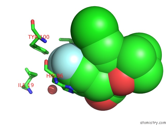
Mono view
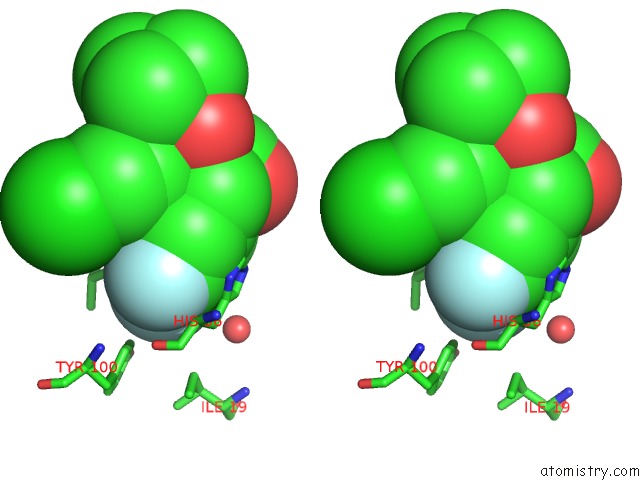
Stereo pair view
Mono view
Stereo pair view
A full contact list of Fluorine with other atoms in the F binding
site number 2 of Exhaustive Fluorine Scanning Towards Potent P53-MDM2 Antagonist within 5.0Å range:
|
Reference:
Y.Huang,
S.Wolf,
D.Koes,
G.M.Popowicz,
C.J.Camacho,
T.A.Holak,
A.Domling.
Exhaustive Fluorine Scanning Toward Potent P53-MDM2 Antagonists. Chemmedchem V. 7 49 2012.
ISSN: ISSN 1860-7179
PubMed: 21954050
DOI: 10.1002/CMDC.201100428
Page generated: Mon Jul 14 19:30:31 2025
ISSN: ISSN 1860-7179
PubMed: 21954050
DOI: 10.1002/CMDC.201100428
Last articles
Zn in 3NDIZn in 3NE8
Zn in 3NDJ
Zn in 3NCJ
Zn in 3NCU
Zn in 3NAC
Zn in 3N9R
Zn in 3NB5
Zn in 3NA9
Zn in 3NAT