Fluorine »
PDB 4b00-4bjk »
4b6s »
Fluorine in PDB 4b6s: Structure of Helicobacter Pylori Type II Dehydroquinase Inhibited By (2S)-2-Perfluorobenzyl-3-Dehydroquinic Acid
Enzymatic activity of Structure of Helicobacter Pylori Type II Dehydroquinase Inhibited By (2S)-2-Perfluorobenzyl-3-Dehydroquinic Acid
All present enzymatic activity of Structure of Helicobacter Pylori Type II Dehydroquinase Inhibited By (2S)-2-Perfluorobenzyl-3-Dehydroquinic Acid:
4.2.1.10;
4.2.1.10;
Protein crystallography data
The structure of Structure of Helicobacter Pylori Type II Dehydroquinase Inhibited By (2S)-2-Perfluorobenzyl-3-Dehydroquinic Acid, PDB code: 4b6s
was solved by
J.M.Otero,
A.L.Llamas-Saiz,
E.Lence,
L.Tizon,
A.Peon,
V.F.V.Prazeres,
H.Lamb,
A.R.Hawkins,
C.Gonzalez-Bello,
M.J.Van Raaij,
with X-Ray Crystallography technique. A brief refinement statistics is given in the table below:
| Resolution Low / High (Å) | 58.65 / 1.90 |
| Space group | P 42 2 2 |
| Cell size a, b, c (Å), α, β, γ (°) | 100.190, 100.190, 104.580, 90.00, 90.00, 90.00 |
| R / Rfree (%) | 22.6 / 27.8 |
Fluorine Binding Sites:
Pages:
>>> Page 1 <<< Page 2, Binding sites: 11 - 15;Binding sites:
The binding sites of Fluorine atom in the Structure of Helicobacter Pylori Type II Dehydroquinase Inhibited By (2S)-2-Perfluorobenzyl-3-Dehydroquinic Acid (pdb code 4b6s). This binding sites where shown within 5.0 Angstroms radius around Fluorine atom.In total 15 binding sites of Fluorine where determined in the Structure of Helicobacter Pylori Type II Dehydroquinase Inhibited By (2S)-2-Perfluorobenzyl-3-Dehydroquinic Acid, PDB code: 4b6s:
Jump to Fluorine binding site number: 1; 2; 3; 4; 5; 6; 7; 8; 9; 10;
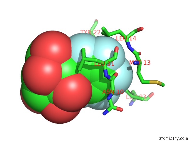
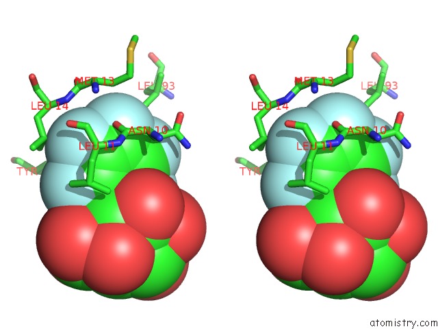
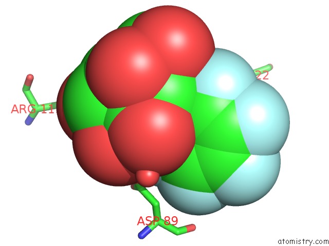
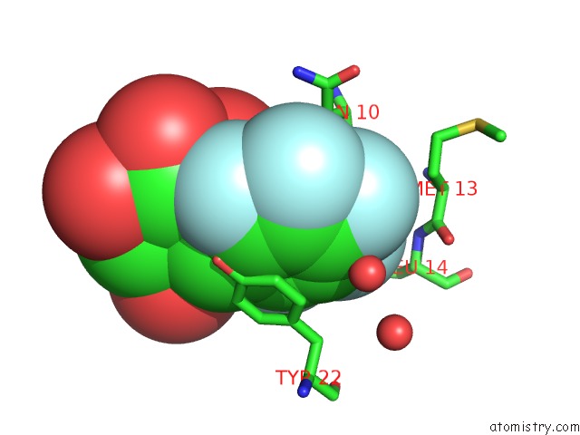
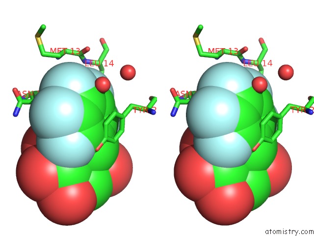
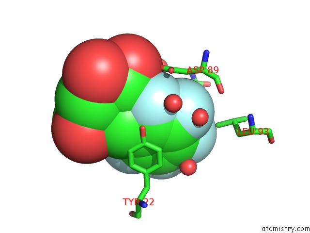
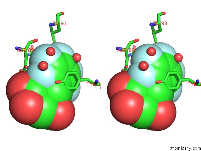
Fluorine binding site 1 out of 15 in 4b6s
Go back to
Fluorine binding site 1 out
of 15 in the Structure of Helicobacter Pylori Type II Dehydroquinase Inhibited By (2S)-2-Perfluorobenzyl-3-Dehydroquinic Acid
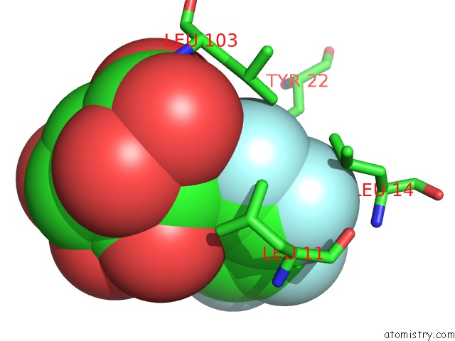
Mono view
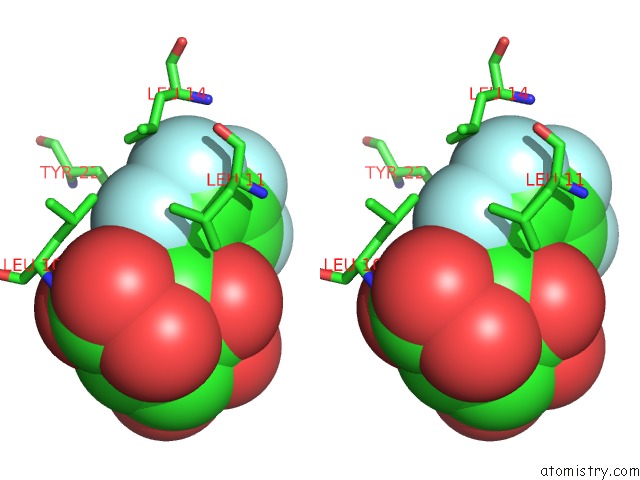
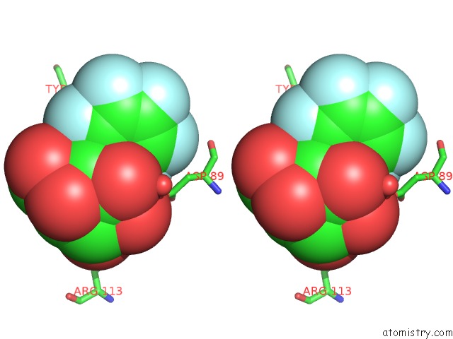
Stereo pair view
Mono view
Stereo pair view
A full contact list of Fluorine with other atoms in the F binding
site number 1 of Structure of Helicobacter Pylori Type II Dehydroquinase Inhibited By (2S)-2-Perfluorobenzyl-3-Dehydroquinic Acid within 5.0Å range:
|
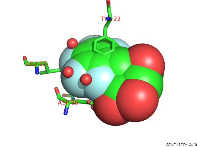
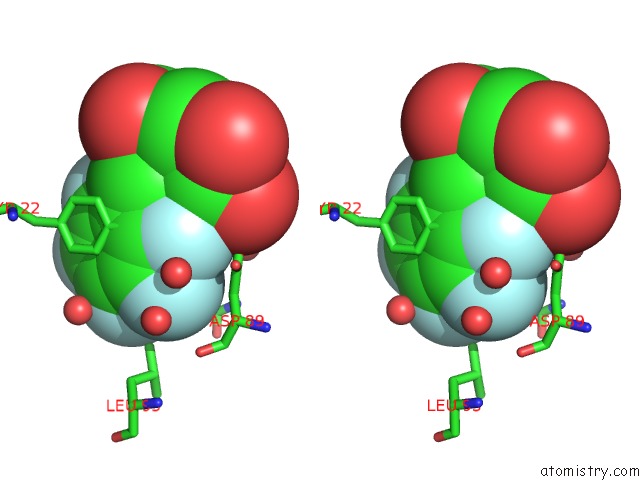
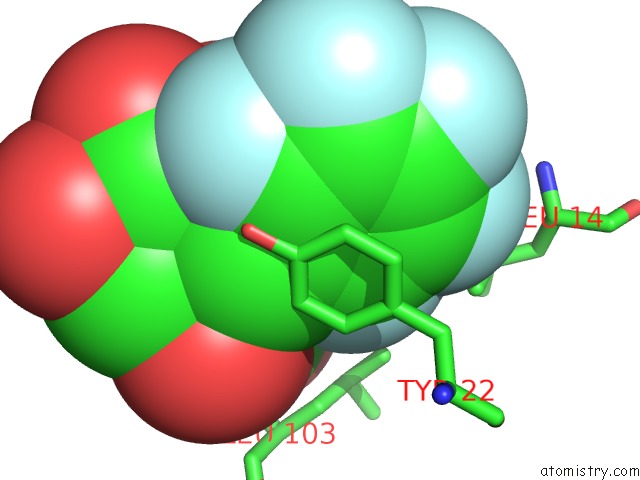
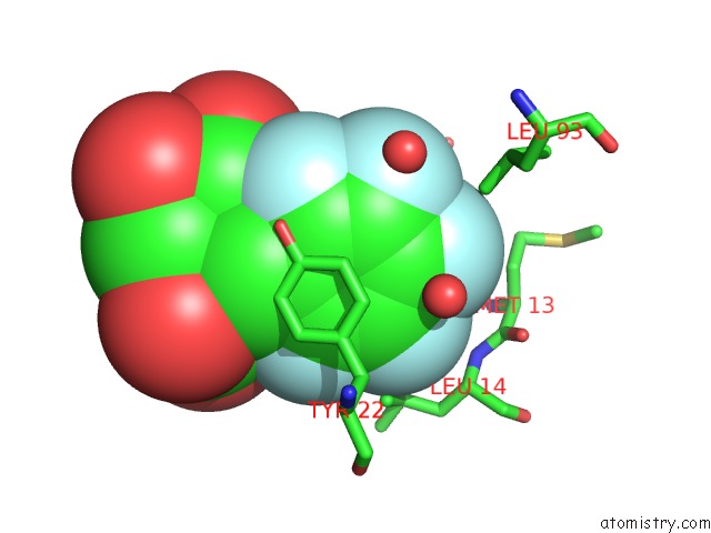
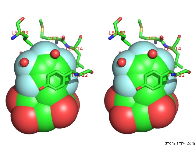
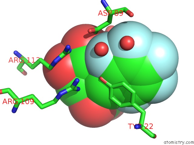
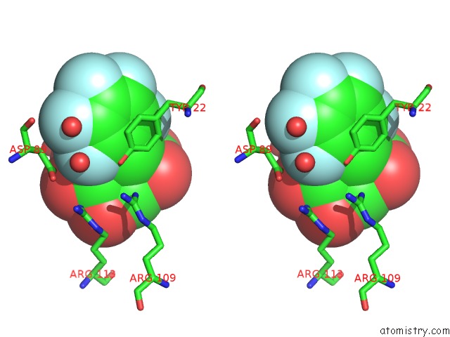
Fluorine binding site 2 out of 15 in 4b6s
Go back to
Fluorine binding site 2 out
of 15 in the Structure of Helicobacter Pylori Type II Dehydroquinase Inhibited By (2S)-2-Perfluorobenzyl-3-Dehydroquinic Acid
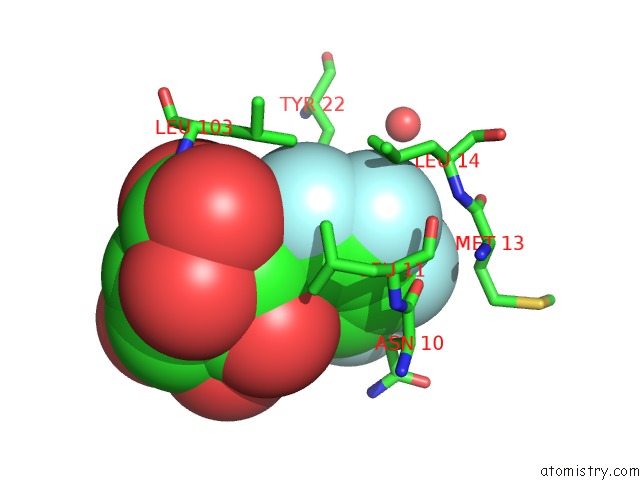
Mono view
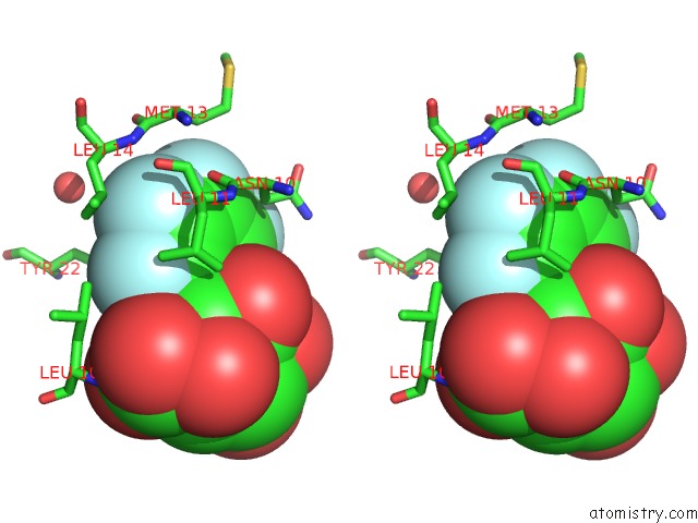
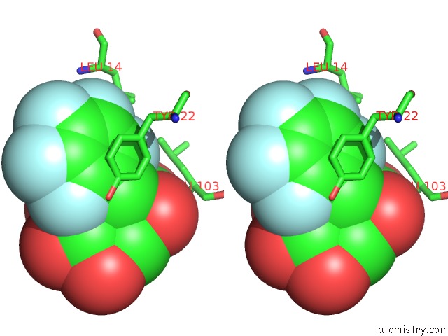
Stereo pair view
Mono view
Stereo pair view
A full contact list of Fluorine with other atoms in the F binding
site number 2 of Structure of Helicobacter Pylori Type II Dehydroquinase Inhibited By (2S)-2-Perfluorobenzyl-3-Dehydroquinic Acid within 5.0Å range:
|
Fluorine binding site 3 out of 15 in 4b6s
Go back to
Fluorine binding site 3 out
of 15 in the Structure of Helicobacter Pylori Type II Dehydroquinase Inhibited By (2S)-2-Perfluorobenzyl-3-Dehydroquinic Acid
Mono view
Stereo pair view
Mono view
Stereo pair view
A full contact list of Fluorine with other atoms in the F binding
site number 3 of Structure of Helicobacter Pylori Type II Dehydroquinase Inhibited By (2S)-2-Perfluorobenzyl-3-Dehydroquinic Acid within 5.0Å range:
|
Fluorine binding site 4 out of 15 in 4b6s
Go back to
Fluorine binding site 4 out
of 15 in the Structure of Helicobacter Pylori Type II Dehydroquinase Inhibited By (2S)-2-Perfluorobenzyl-3-Dehydroquinic Acid
Mono view
Stereo pair view
Mono view
Stereo pair view
A full contact list of Fluorine with other atoms in the F binding
site number 4 of Structure of Helicobacter Pylori Type II Dehydroquinase Inhibited By (2S)-2-Perfluorobenzyl-3-Dehydroquinic Acid within 5.0Å range:
|
Fluorine binding site 5 out of 15 in 4b6s
Go back to
Fluorine binding site 5 out
of 15 in the Structure of Helicobacter Pylori Type II Dehydroquinase Inhibited By (2S)-2-Perfluorobenzyl-3-Dehydroquinic Acid
Mono view
Stereo pair view
Mono view
Stereo pair view
A full contact list of Fluorine with other atoms in the F binding
site number 5 of Structure of Helicobacter Pylori Type II Dehydroquinase Inhibited By (2S)-2-Perfluorobenzyl-3-Dehydroquinic Acid within 5.0Å range:
|
Fluorine binding site 6 out of 15 in 4b6s
Go back to
Fluorine binding site 6 out
of 15 in the Structure of Helicobacter Pylori Type II Dehydroquinase Inhibited By (2S)-2-Perfluorobenzyl-3-Dehydroquinic Acid
Mono view
Stereo pair view
Mono view
Stereo pair view
A full contact list of Fluorine with other atoms in the F binding
site number 6 of Structure of Helicobacter Pylori Type II Dehydroquinase Inhibited By (2S)-2-Perfluorobenzyl-3-Dehydroquinic Acid within 5.0Å range:
|
Fluorine binding site 7 out of 15 in 4b6s
Go back to
Fluorine binding site 7 out
of 15 in the Structure of Helicobacter Pylori Type II Dehydroquinase Inhibited By (2S)-2-Perfluorobenzyl-3-Dehydroquinic Acid
Mono view
Stereo pair view
Mono view
Stereo pair view
A full contact list of Fluorine with other atoms in the F binding
site number 7 of Structure of Helicobacter Pylori Type II Dehydroquinase Inhibited By (2S)-2-Perfluorobenzyl-3-Dehydroquinic Acid within 5.0Å range:
|
Fluorine binding site 8 out of 15 in 4b6s
Go back to
Fluorine binding site 8 out
of 15 in the Structure of Helicobacter Pylori Type II Dehydroquinase Inhibited By (2S)-2-Perfluorobenzyl-3-Dehydroquinic Acid
Mono view
Stereo pair view
Mono view
Stereo pair view
A full contact list of Fluorine with other atoms in the F binding
site number 8 of Structure of Helicobacter Pylori Type II Dehydroquinase Inhibited By (2S)-2-Perfluorobenzyl-3-Dehydroquinic Acid within 5.0Å range:
|
Fluorine binding site 9 out of 15 in 4b6s
Go back to
Fluorine binding site 9 out
of 15 in the Structure of Helicobacter Pylori Type II Dehydroquinase Inhibited By (2S)-2-Perfluorobenzyl-3-Dehydroquinic Acid
Mono view
Stereo pair view
Mono view
Stereo pair view
A full contact list of Fluorine with other atoms in the F binding
site number 9 of Structure of Helicobacter Pylori Type II Dehydroquinase Inhibited By (2S)-2-Perfluorobenzyl-3-Dehydroquinic Acid within 5.0Å range:
|
Fluorine binding site 10 out of 15 in 4b6s
Go back to
Fluorine binding site 10 out
of 15 in the Structure of Helicobacter Pylori Type II Dehydroquinase Inhibited By (2S)-2-Perfluorobenzyl-3-Dehydroquinic Acid
Mono view
Stereo pair view
Mono view
Stereo pair view
A full contact list of Fluorine with other atoms in the F binding
site number 10 of Structure of Helicobacter Pylori Type II Dehydroquinase Inhibited By (2S)-2-Perfluorobenzyl-3-Dehydroquinic Acid within 5.0Å range:
|
Reference:
E.Lence,
L.Tizon,
J.M.Otero,
A.Peon,
V.F.Prazeres,
A.L.Llamas-Saiz,
G.C.Fox,
M.J.Van Raaij,
H.Lamb,
A.R.Hawkins,
C.Gonzalez-Bello.
Mechanistic Basis of the Inhibition of Type II Dehydroquinase By (2S)- and (2R)-2-Benzyl-3-Dehydroquinic Acids. Acs Chem. Biol. V. 8 568 2013.
ISSN: ESSN 1554-8937
PubMed: 23198883
DOI: 10.1021/CB300493S
Page generated: Mon Jul 14 20:30:41 2025
ISSN: ESSN 1554-8937
PubMed: 23198883
DOI: 10.1021/CB300493S
Last articles
Fe in 7W4LFe in 7W4K
Fe in 7W4J
Fe in 7W4G
Fe in 7W4F
Fe in 7W4E
Fe in 7W4D
Fe in 7W4C
Fe in 7W35
Fe in 7W3E