Fluorine »
PDB 4b00-4bjk »
4bb0 »
Fluorine in PDB 4bb0: Structure of A Putative Epoxide Hydrolase Q244E Mutant From Pseudomonas Aeruginosa, with Bound Mfa.
Protein crystallography data
The structure of Structure of A Putative Epoxide Hydrolase Q244E Mutant From Pseudomonas Aeruginosa, with Bound Mfa., PDB code: 4bb0
was solved by
J.W.Schmidberger,
R.Schnell,
G.Schneider,
with X-Ray Crystallography technique. A brief refinement statistics is given in the table below:
| Resolution Low / High (Å) | 27.16 / 1.77 |
| Space group | P 41 21 2 |
| Cell size a, b, c (Å), α, β, γ (°) | 83.242, 83.242, 140.541, 90.00, 90.00, 90.00 |
| R / Rfree (%) | 15.092 / 21.522 |
Fluorine Binding Sites:
The binding sites of Fluorine atom in the Structure of A Putative Epoxide Hydrolase Q244E Mutant From Pseudomonas Aeruginosa, with Bound Mfa.
(pdb code 4bb0). This binding sites where shown within
5.0 Angstroms radius around Fluorine atom.
In total 5 binding sites of Fluorine where determined in the Structure of A Putative Epoxide Hydrolase Q244E Mutant From Pseudomonas Aeruginosa, with Bound Mfa., PDB code: 4bb0:
Jump to Fluorine binding site number: 1; 2; 3; 4; 5;
In total 5 binding sites of Fluorine where determined in the Structure of A Putative Epoxide Hydrolase Q244E Mutant From Pseudomonas Aeruginosa, with Bound Mfa., PDB code: 4bb0:
Jump to Fluorine binding site number: 1; 2; 3; 4; 5;
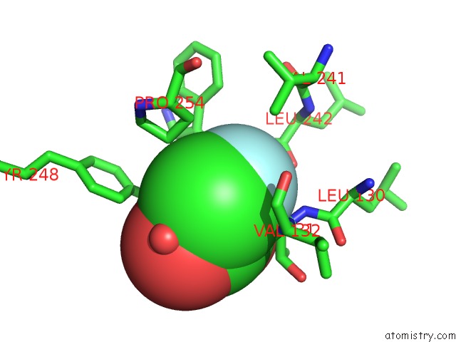
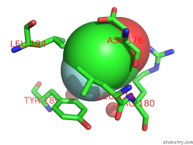
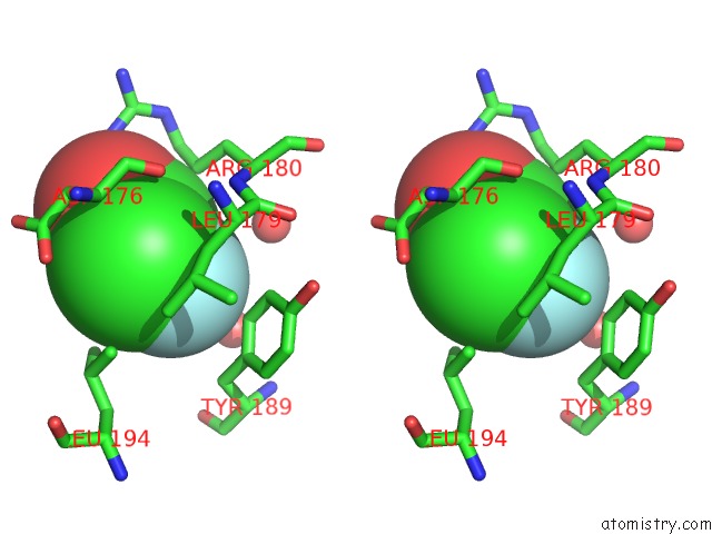
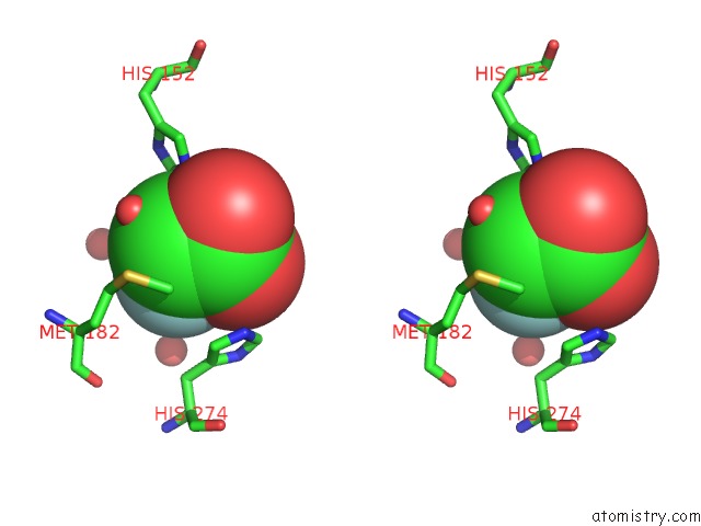
Fluorine binding site 1 out of 5 in 4bb0
Go back to
Fluorine binding site 1 out
of 5 in the Structure of A Putative Epoxide Hydrolase Q244E Mutant From Pseudomonas Aeruginosa, with Bound Mfa.
Mono view
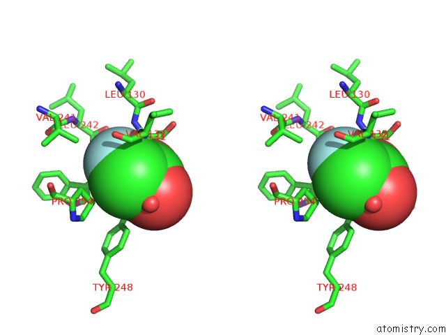
Stereo pair view
Mono view
Stereo pair view
A full contact list of Fluorine with other atoms in the F binding
site number 1 of Structure of A Putative Epoxide Hydrolase Q244E Mutant From Pseudomonas Aeruginosa, with Bound Mfa. within 5.0Å range:
|
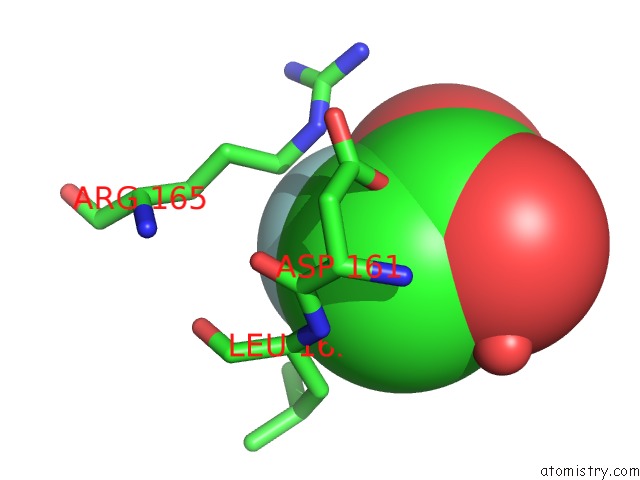
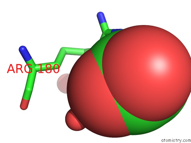
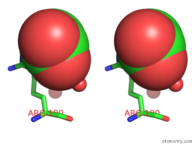
Fluorine binding site 2 out of 5 in 4bb0
Go back to
Fluorine binding site 2 out
of 5 in the Structure of A Putative Epoxide Hydrolase Q244E Mutant From Pseudomonas Aeruginosa, with Bound Mfa.
Mono view
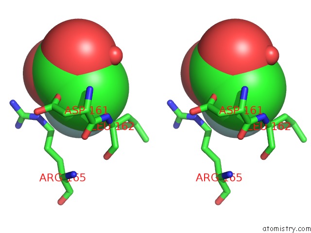
Stereo pair view
Mono view
Stereo pair view
A full contact list of Fluorine with other atoms in the F binding
site number 2 of Structure of A Putative Epoxide Hydrolase Q244E Mutant From Pseudomonas Aeruginosa, with Bound Mfa. within 5.0Å range:
|
Fluorine binding site 3 out of 5 in 4bb0
Go back to
Fluorine binding site 3 out
of 5 in the Structure of A Putative Epoxide Hydrolase Q244E Mutant From Pseudomonas Aeruginosa, with Bound Mfa.
Mono view
Stereo pair view
Mono view
Stereo pair view
A full contact list of Fluorine with other atoms in the F binding
site number 3 of Structure of A Putative Epoxide Hydrolase Q244E Mutant From Pseudomonas Aeruginosa, with Bound Mfa. within 5.0Å range:
|
Fluorine binding site 4 out of 5 in 4bb0
Go back to
Fluorine binding site 4 out
of 5 in the Structure of A Putative Epoxide Hydrolase Q244E Mutant From Pseudomonas Aeruginosa, with Bound Mfa.
Mono view
Stereo pair view
Mono view
Stereo pair view
A full contact list of Fluorine with other atoms in the F binding
site number 4 of Structure of A Putative Epoxide Hydrolase Q244E Mutant From Pseudomonas Aeruginosa, with Bound Mfa. within 5.0Å range:
|
Fluorine binding site 5 out of 5 in 4bb0
Go back to
Fluorine binding site 5 out
of 5 in the Structure of A Putative Epoxide Hydrolase Q244E Mutant From Pseudomonas Aeruginosa, with Bound Mfa.

Mono view
Stereo pair view
Mono view
Stereo pair view
A full contact list of Fluorine with other atoms in the F binding
site number 5 of Structure of A Putative Epoxide Hydrolase Q244E Mutant From Pseudomonas Aeruginosa, with Bound Mfa. within 5.0Å range:
|
Reference:
J.W.Schmidberger,
R.Schnell,
G.Schneider.
Structure of A Putative Epoxide Hydrolase Mutant To Be Published.
Page generated: Mon Jul 14 20:33:21 2025
Last articles
Fe in 7W4FFe in 7W4E
Fe in 7W4D
Fe in 7W4C
Fe in 7W35
Fe in 7W3E
Fe in 7W32
Fe in 7W2Y
Fe in 7W31
Fe in 7W2U