Fluorine »
PDB 4e3n-4f9m »
4ear »
Fluorine in PDB 4ear: Crystal Structure of Purine Nucleoside Phosphorylase (W16Y, W94Y, W178Y, H257W) Mutant From Human Complexed with Dadme-Immg and Phosphate
Enzymatic activity of Crystal Structure of Purine Nucleoside Phosphorylase (W16Y, W94Y, W178Y, H257W) Mutant From Human Complexed with Dadme-Immg and Phosphate
All present enzymatic activity of Crystal Structure of Purine Nucleoside Phosphorylase (W16Y, W94Y, W178Y, H257W) Mutant From Human Complexed with Dadme-Immg and Phosphate:
2.4.2.1;
2.4.2.1;
Protein crystallography data
The structure of Crystal Structure of Purine Nucleoside Phosphorylase (W16Y, W94Y, W178Y, H257W) Mutant From Human Complexed with Dadme-Immg and Phosphate, PDB code: 4ear
was solved by
A.M.Haapalainen,
M.C.Ho,
J.J.Suarez,
S.C.Almo,
V.L.Schramm,
with X-Ray Crystallography technique. A brief refinement statistics is given in the table below:
| Resolution Low / High (Å) | 41.24 / 1.70 |
| Space group | P 21 21 21 |
| Cell size a, b, c (Å), α, β, γ (°) | 55.916, 131.100, 137.680, 90.00, 90.00, 90.00 |
| R / Rfree (%) | 17.5 / 20.3 |
Fluorine Binding Sites:
The binding sites of Fluorine atom in the Crystal Structure of Purine Nucleoside Phosphorylase (W16Y, W94Y, W178Y, H257W) Mutant From Human Complexed with Dadme-Immg and Phosphate
(pdb code 4ear). This binding sites where shown within
5.0 Angstroms radius around Fluorine atom.
In total 3 binding sites of Fluorine where determined in the Crystal Structure of Purine Nucleoside Phosphorylase (W16Y, W94Y, W178Y, H257W) Mutant From Human Complexed with Dadme-Immg and Phosphate, PDB code: 4ear:
Jump to Fluorine binding site number: 1; 2; 3;
In total 3 binding sites of Fluorine where determined in the Crystal Structure of Purine Nucleoside Phosphorylase (W16Y, W94Y, W178Y, H257W) Mutant From Human Complexed with Dadme-Immg and Phosphate, PDB code: 4ear:
Jump to Fluorine binding site number: 1; 2; 3;
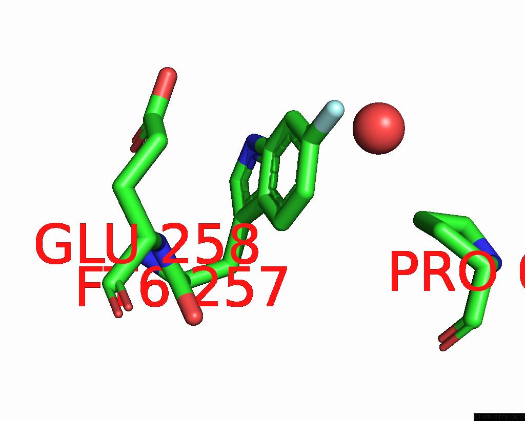
Fluorine binding site 1 out of 3 in 4ear
Go back to
Fluorine binding site 1 out
of 3 in the Crystal Structure of Purine Nucleoside Phosphorylase (W16Y, W94Y, W178Y, H257W) Mutant From Human Complexed with Dadme-Immg and Phosphate

Mono view

Stereo pair view
Mono view
Stereo pair view
A full contact list of Fluorine with other atoms in the F binding
site number 1 of Crystal Structure of Purine Nucleoside Phosphorylase (W16Y, W94Y, W178Y, H257W) Mutant From Human Complexed with Dadme-Immg and Phosphate within 5.0Å range:
|
Fluorine binding site 2 out of 3 in 4ear
Go back to
Fluorine binding site 2 out
of 3 in the Crystal Structure of Purine Nucleoside Phosphorylase (W16Y, W94Y, W178Y, H257W) Mutant From Human Complexed with Dadme-Immg and Phosphate
Mono view

Stereo pair view
Mono view
Stereo pair view
A full contact list of Fluorine with other atoms in the F binding
site number 2 of Crystal Structure of Purine Nucleoside Phosphorylase (W16Y, W94Y, W178Y, H257W) Mutant From Human Complexed with Dadme-Immg and Phosphate within 5.0Å range:
|
Fluorine binding site 3 out of 3 in 4ear
Go back to
Fluorine binding site 3 out
of 3 in the Crystal Structure of Purine Nucleoside Phosphorylase (W16Y, W94Y, W178Y, H257W) Mutant From Human Complexed with Dadme-Immg and Phosphate

Mono view

Stereo pair view
Mono view
Stereo pair view
A full contact list of Fluorine with other atoms in the F binding
site number 3 of Crystal Structure of Purine Nucleoside Phosphorylase (W16Y, W94Y, W178Y, H257W) Mutant From Human Complexed with Dadme-Immg and Phosphate within 5.0Å range:
|
Reference:
J.Suarez,
A.M.Haapalainen,
S.M.Cahill,
M.C.Ho,
F.Yan,
S.C.Almo,
V.L.Schramm.
Catalytic Site Conformations in Human Pnp By (19)F-uc(Nmr) and Crystallography. Chem.Biol. V. 20 212 2013.
ISSN: ISSN 1074-5521
PubMed: 23438750
DOI: 10.1016/J.CHEMBIOL.2013.01.009
Page generated: Mon Jul 14 21:23:56 2025
ISSN: ISSN 1074-5521
PubMed: 23438750
DOI: 10.1016/J.CHEMBIOL.2013.01.009
Last articles
Mg in 8VSDMg in 8VQV
Mg in 8VQI
Mg in 8VPB
Mg in 8VOX
Mg in 8VOW
Mg in 8VOI
Mg in 8VOA
Mg in 8VO9
Mg in 8VO8