Fluorine »
PDB 4kbk-4l3l »
4l3l »
Fluorine in PDB 4l3l: Crystal Structures of Human P70S6K1 Kinase Domain (Zinc Anomalous)
Protein crystallography data
The structure of Crystal Structures of Human P70S6K1 Kinase Domain (Zinc Anomalous), PDB code: 4l3l
was solved by
J.Wang,
C.Zhong,
J.Ding,
with X-Ray Crystallography technique. A brief refinement statistics is given in the table below:
| Resolution Low / High (Å) | 46.48 / 2.10 |
| Space group | P 41 21 2 |
| Cell size a, b, c (Å), α, β, γ (°) | 69.408, 69.408, 143.378, 90.00, 90.00, 90.00 |
| R / Rfree (%) | 20.7 / 24.7 |
Other elements in 4l3l:
The structure of Crystal Structures of Human P70S6K1 Kinase Domain (Zinc Anomalous) also contains other interesting chemical elements:
| Zinc | (Zn) | 1 atom |
Fluorine Binding Sites:
The binding sites of Fluorine atom in the Crystal Structures of Human P70S6K1 Kinase Domain (Zinc Anomalous)
(pdb code 4l3l). This binding sites where shown within
5.0 Angstroms radius around Fluorine atom.
In total 3 binding sites of Fluorine where determined in the Crystal Structures of Human P70S6K1 Kinase Domain (Zinc Anomalous), PDB code: 4l3l:
Jump to Fluorine binding site number: 1; 2; 3;
In total 3 binding sites of Fluorine where determined in the Crystal Structures of Human P70S6K1 Kinase Domain (Zinc Anomalous), PDB code: 4l3l:
Jump to Fluorine binding site number: 1; 2; 3;
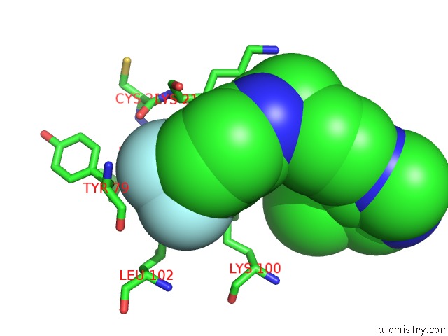
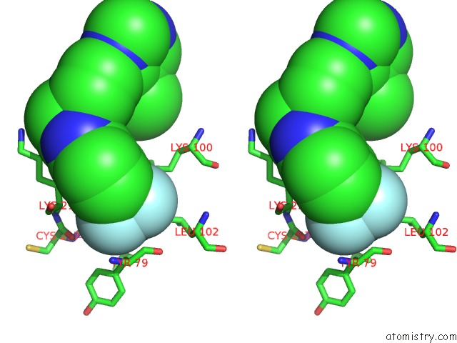
Fluorine binding site 1 out of 3 in 4l3l
Go back to
Fluorine binding site 1 out
of 3 in the Crystal Structures of Human P70S6K1 Kinase Domain (Zinc Anomalous)
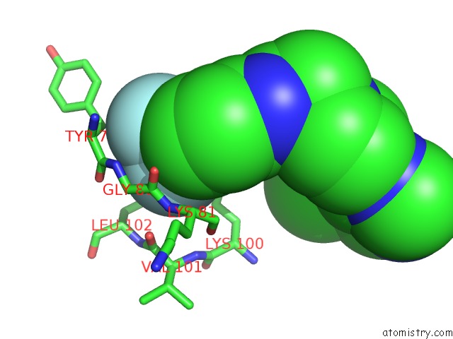
Mono view
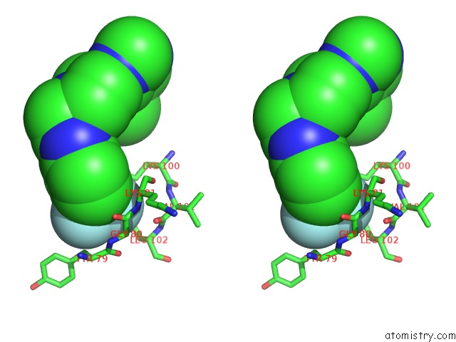
Stereo pair view
Mono view
Stereo pair view
A full contact list of Fluorine with other atoms in the F binding
site number 1 of Crystal Structures of Human P70S6K1 Kinase Domain (Zinc Anomalous) within 5.0Å range:
|
Fluorine binding site 2 out of 3 in 4l3l
Go back to
Fluorine binding site 2 out
of 3 in the Crystal Structures of Human P70S6K1 Kinase Domain (Zinc Anomalous)
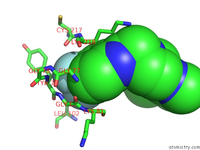
Mono view
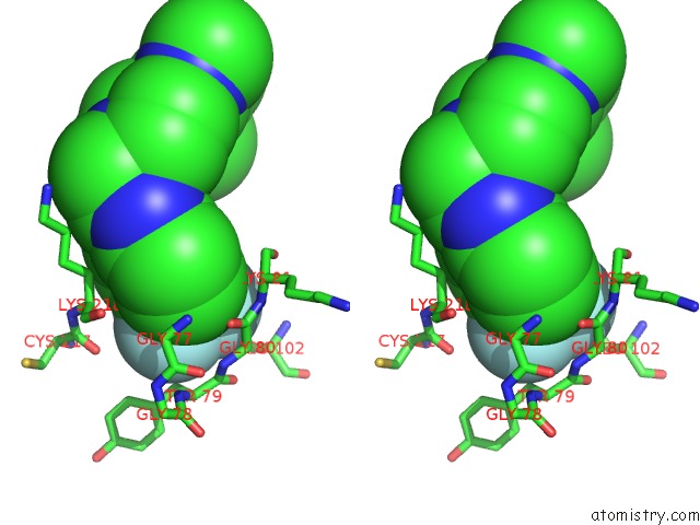
Stereo pair view
Mono view
Stereo pair view
A full contact list of Fluorine with other atoms in the F binding
site number 2 of Crystal Structures of Human P70S6K1 Kinase Domain (Zinc Anomalous) within 5.0Å range:
|
Fluorine binding site 3 out of 3 in 4l3l
Go back to
Fluorine binding site 3 out
of 3 in the Crystal Structures of Human P70S6K1 Kinase Domain (Zinc Anomalous)
Mono view
Stereo pair view
Mono view
Stereo pair view
A full contact list of Fluorine with other atoms in the F binding
site number 3 of Crystal Structures of Human P70S6K1 Kinase Domain (Zinc Anomalous) within 5.0Å range:
|
Reference:
J.Wang,
C.Zhong,
F.Wang,
F.Qu,
J.Ding.
Crystal Structures of S6K1 Provide Insights Into the Regulation Mechanism of S6K1 By the Hydrophobic Motif Biochem.J. V. 454 39 2013.
ISSN: ISSN 0264-6021
PubMed: 23731517
DOI: 10.1042/BJ20121863
Page generated: Mon Jul 14 22:57:12 2025
ISSN: ISSN 0264-6021
PubMed: 23731517
DOI: 10.1042/BJ20121863
Last articles
Mg in 1FIRMg in 1FI1
Mg in 1FHV
Mg in 1FGS
Mg in 1FEZ
Mg in 1FFK
Mg in 1FFH
Mg in 1FC5
Mg in 1FDG
Mg in 1FD5