Fluorine »
PDB 4lvt-4mm8 »
4lxi »
Fluorine in PDB 4lxi: Crystal Structure of the S105A Mutant of A Carbon-Carbon Bond Hydrolase, DXNB2 From Sphingomonas Wittichii RW1, in Complex with 5, 8-Dif Hopda
Enzymatic activity of Crystal Structure of the S105A Mutant of A Carbon-Carbon Bond Hydrolase, DXNB2 From Sphingomonas Wittichii RW1, in Complex with 5, 8-Dif Hopda
All present enzymatic activity of Crystal Structure of the S105A Mutant of A Carbon-Carbon Bond Hydrolase, DXNB2 From Sphingomonas Wittichii RW1, in Complex with 5, 8-Dif Hopda:
3.7.1.8;
3.7.1.8;
Protein crystallography data
The structure of Crystal Structure of the S105A Mutant of A Carbon-Carbon Bond Hydrolase, DXNB2 From Sphingomonas Wittichii RW1, in Complex with 5, 8-Dif Hopda, PDB code: 4lxi
was solved by
S.Bhowmik,
J.T.Bolin,
with X-Ray Crystallography technique. A brief refinement statistics is given in the table below:
| Resolution Low / High (Å) | 57.26 / 2.17 |
| Space group | P 65 2 2 |
| Cell size a, b, c (Å), α, β, γ (°) | 66.155, 66.155, 342.180, 90.00, 90.00, 120.00 |
| R / Rfree (%) | 20.4 / 25.8 |
Other elements in 4lxi:
The structure of Crystal Structure of the S105A Mutant of A Carbon-Carbon Bond Hydrolase, DXNB2 From Sphingomonas Wittichii RW1, in Complex with 5, 8-Dif Hopda also contains other interesting chemical elements:
| Sodium | (Na) | 1 atom |
Fluorine Binding Sites:
The binding sites of Fluorine atom in the Crystal Structure of the S105A Mutant of A Carbon-Carbon Bond Hydrolase, DXNB2 From Sphingomonas Wittichii RW1, in Complex with 5, 8-Dif Hopda
(pdb code 4lxi). This binding sites where shown within
5.0 Angstroms radius around Fluorine atom.
In total 2 binding sites of Fluorine where determined in the Crystal Structure of the S105A Mutant of A Carbon-Carbon Bond Hydrolase, DXNB2 From Sphingomonas Wittichii RW1, in Complex with 5, 8-Dif Hopda, PDB code: 4lxi:
Jump to Fluorine binding site number: 1; 2;
In total 2 binding sites of Fluorine where determined in the Crystal Structure of the S105A Mutant of A Carbon-Carbon Bond Hydrolase, DXNB2 From Sphingomonas Wittichii RW1, in Complex with 5, 8-Dif Hopda, PDB code: 4lxi:
Jump to Fluorine binding site number: 1; 2;
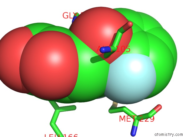
Fluorine binding site 1 out of 2 in 4lxi
Go back to
Fluorine binding site 1 out
of 2 in the Crystal Structure of the S105A Mutant of A Carbon-Carbon Bond Hydrolase, DXNB2 From Sphingomonas Wittichii RW1, in Complex with 5, 8-Dif Hopda
Mono view

Stereo pair view
Mono view
Stereo pair view
A full contact list of Fluorine with other atoms in the F binding
site number 1 of Crystal Structure of the S105A Mutant of A Carbon-Carbon Bond Hydrolase, DXNB2 From Sphingomonas Wittichii RW1, in Complex with 5, 8-Dif Hopda within 5.0Å range:
|
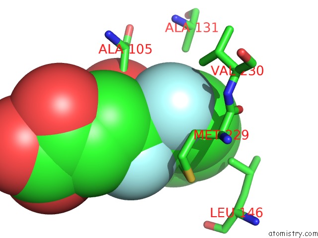
Fluorine binding site 2 out of 2 in 4lxi
Go back to
Fluorine binding site 2 out
of 2 in the Crystal Structure of the S105A Mutant of A Carbon-Carbon Bond Hydrolase, DXNB2 From Sphingomonas Wittichii RW1, in Complex with 5, 8-Dif Hopda
Mono view

Stereo pair view
Mono view
Stereo pair view
A full contact list of Fluorine with other atoms in the F binding
site number 2 of Crystal Structure of the S105A Mutant of A Carbon-Carbon Bond Hydrolase, DXNB2 From Sphingomonas Wittichii RW1, in Complex with 5, 8-Dif Hopda within 5.0Å range:
|
Reference:
A.C.Ruzzini,
S.Bhowmik,
S.Ghosh,
K.C.Yam,
J.T.Bolin,
L.D.Eltis.
A Substrate-Assisted Mechanism of Nucleophile Activation in A Ser-His-Asp Containing C-C Bond Hydrolase. Biochemistry V. 52 7428 2013.
ISSN: ISSN 0006-2960
PubMed: 24067021
DOI: 10.1021/BI401156A
Page generated: Mon Jul 14 23:06:46 2025
ISSN: ISSN 0006-2960
PubMed: 24067021
DOI: 10.1021/BI401156A
Last articles
K in 7PWYK in 7PXE
K in 7PXF
K in 7PVC
K in 7PQU
K in 7PQT
K in 7PS8
K in 7PV8
K in 7POZ
K in 7PNL