Fluorine »
PDB 4lvt-4mm8 »
4mk0 »
Fluorine in PDB 4mk0: Crystal Structure of G Protein-Coupled Receptor Kinase 2 in Complex with A A Rationally Designed Paroxetine Derivative
Enzymatic activity of Crystal Structure of G Protein-Coupled Receptor Kinase 2 in Complex with A A Rationally Designed Paroxetine Derivative
All present enzymatic activity of Crystal Structure of G Protein-Coupled Receptor Kinase 2 in Complex with A A Rationally Designed Paroxetine Derivative:
2.7.11.15;
2.7.11.15;
Protein crystallography data
The structure of Crystal Structure of G Protein-Coupled Receptor Kinase 2 in Complex with A A Rationally Designed Paroxetine Derivative, PDB code: 4mk0
was solved by
K.T.Homan,
J.J.G.Tesmer,
with X-Ray Crystallography technique. A brief refinement statistics is given in the table below:
| Resolution Low / High (Å) | 19.99 / 2.40 |
| Space group | C 2 2 21 |
| Cell size a, b, c (Å), α, β, γ (°) | 61.222, 240.973, 212.083, 90.00, 90.00, 90.00 |
| R / Rfree (%) | 17.5 / 23.8 |
Fluorine Binding Sites:
The binding sites of Fluorine atom in the Crystal Structure of G Protein-Coupled Receptor Kinase 2 in Complex with A A Rationally Designed Paroxetine Derivative
(pdb code 4mk0). This binding sites where shown within
5.0 Angstroms radius around Fluorine atom.
In total only one binding site of Fluorine was determined in the Crystal Structure of G Protein-Coupled Receptor Kinase 2 in Complex with A A Rationally Designed Paroxetine Derivative, PDB code: 4mk0:
In total only one binding site of Fluorine was determined in the Crystal Structure of G Protein-Coupled Receptor Kinase 2 in Complex with A A Rationally Designed Paroxetine Derivative, PDB code: 4mk0:
Fluorine binding site 1 out of 1 in 4mk0
Go back to
Fluorine binding site 1 out
of 1 in the Crystal Structure of G Protein-Coupled Receptor Kinase 2 in Complex with A A Rationally Designed Paroxetine Derivative
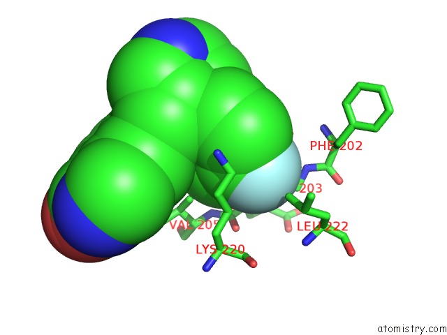
Mono view
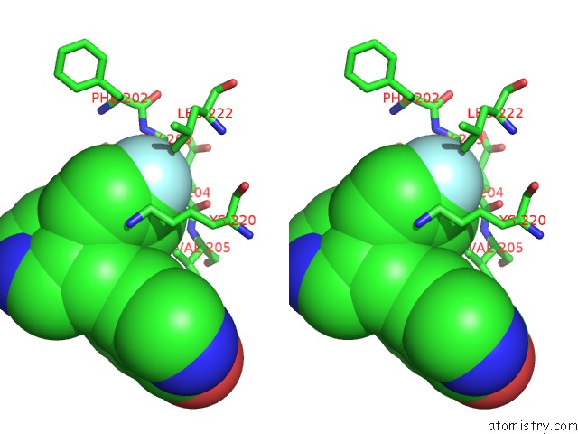
Stereo pair view
Mono view
Stereo pair view
A full contact list of Fluorine with other atoms in the F binding
site number 1 of Crystal Structure of G Protein-Coupled Receptor Kinase 2 in Complex with A A Rationally Designed Paroxetine Derivative within 5.0Å range:
|
Reference:
K.T.Homan,
E.Wu,
M.W.Wilson,
P.Singh,
S.D.Larsen,
J.J.Tesmer.
Structural and Functional Analysis of G Protein-Coupled Receptor Kinase Inhibition By Paroxetine and A Rationally Designed Analog. Mol.Pharmacol. V. 85 237 2014.
ISSN: ISSN 0026-895X
PubMed: 24220010
DOI: 10.1124/MOL.113.089631
Page generated: Mon Jul 14 23:19:52 2025
ISSN: ISSN 0026-895X
PubMed: 24220010
DOI: 10.1124/MOL.113.089631
Last articles
Mg in 1XMQMg in 1XOM
Mg in 1XMO
Mg in 1XNG
Mg in 1XN0
Mg in 1XMY
Mg in 1XMV
Mg in 1XMX
Mg in 1XMI
Mg in 1XMU