Fluorine »
PDB 4mm9-4ncg »
4nam »
Fluorine in PDB 4nam: 1.7A Structure of 5-Fluoro Tryptophan Labeled Protective Antigen (W206Y)
Protein crystallography data
The structure of 1.7A Structure of 5-Fluoro Tryptophan Labeled Protective Antigen (W206Y), PDB code: 4nam
was solved by
S.Lovell,
K.P.Battaile,
F.Chadegani,
V.Mulangi,
M.Miyagi,
J.G.Bann,
with X-Ray Crystallography technique. A brief refinement statistics is given in the table below:
| Resolution Low / High (Å) | 46.98 / 1.70 |
| Space group | P 21 21 21 |
| Cell size a, b, c (Å), α, β, γ (°) | 71.298, 93.954, 117.727, 90.00, 90.00, 90.00 |
| R / Rfree (%) | 17.9 / 20.8 |
Other elements in 4nam:
The structure of 1.7A Structure of 5-Fluoro Tryptophan Labeled Protective Antigen (W206Y) also contains other interesting chemical elements:
| Calcium | (Ca) | 2 atoms |
Fluorine Binding Sites:
The binding sites of Fluorine atom in the 1.7A Structure of 5-Fluoro Tryptophan Labeled Protective Antigen (W206Y)
(pdb code 4nam). This binding sites where shown within
5.0 Angstroms radius around Fluorine atom.
In total 6 binding sites of Fluorine where determined in the 1.7A Structure of 5-Fluoro Tryptophan Labeled Protective Antigen (W206Y), PDB code: 4nam:
Jump to Fluorine binding site number: 1; 2; 3; 4; 5; 6;
In total 6 binding sites of Fluorine where determined in the 1.7A Structure of 5-Fluoro Tryptophan Labeled Protective Antigen (W206Y), PDB code: 4nam:
Jump to Fluorine binding site number: 1; 2; 3; 4; 5; 6;
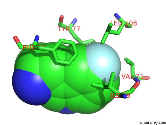
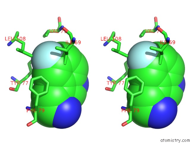
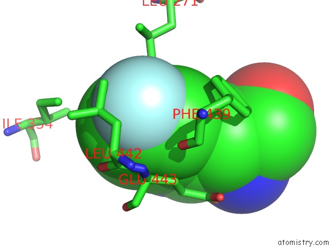
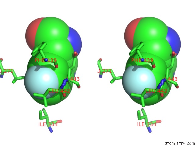
Fluorine binding site 1 out of 6 in 4nam
Go back to
Fluorine binding site 1 out
of 6 in the 1.7A Structure of 5-Fluoro Tryptophan Labeled Protective Antigen (W206Y)
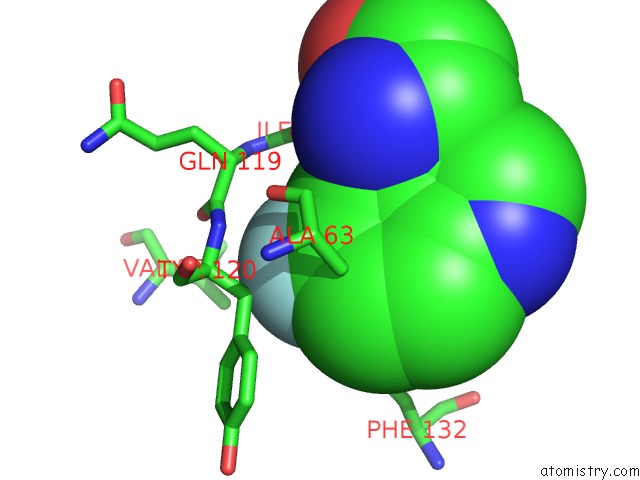
Mono view
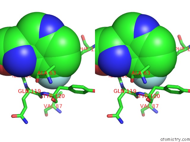
Stereo pair view
Mono view
Stereo pair view
A full contact list of Fluorine with other atoms in the F binding
site number 1 of 1.7A Structure of 5-Fluoro Tryptophan Labeled Protective Antigen (W206Y) within 5.0Å range:
|
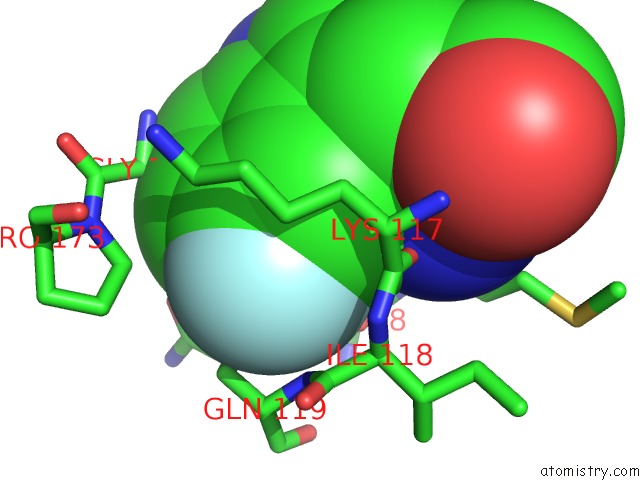
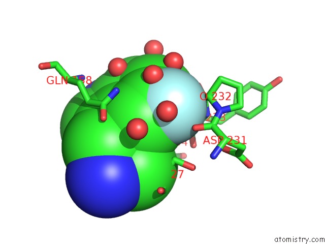
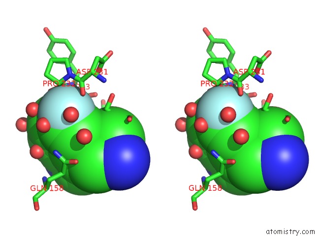
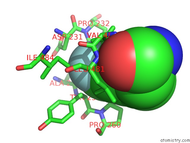
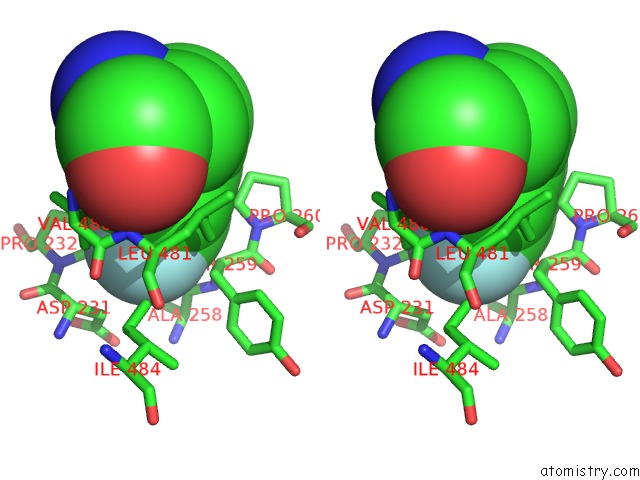
Fluorine binding site 2 out of 6 in 4nam
Go back to
Fluorine binding site 2 out
of 6 in the 1.7A Structure of 5-Fluoro Tryptophan Labeled Protective Antigen (W206Y)
Mono view
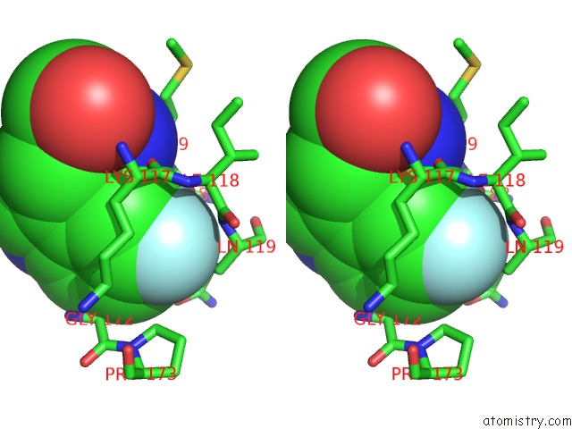
Stereo pair view
Mono view
Stereo pair view
A full contact list of Fluorine with other atoms in the F binding
site number 2 of 1.7A Structure of 5-Fluoro Tryptophan Labeled Protective Antigen (W206Y) within 5.0Å range:
|
Fluorine binding site 3 out of 6 in 4nam
Go back to
Fluorine binding site 3 out
of 6 in the 1.7A Structure of 5-Fluoro Tryptophan Labeled Protective Antigen (W206Y)
Mono view
Stereo pair view
Mono view
Stereo pair view
A full contact list of Fluorine with other atoms in the F binding
site number 3 of 1.7A Structure of 5-Fluoro Tryptophan Labeled Protective Antigen (W206Y) within 5.0Å range:
|
Fluorine binding site 4 out of 6 in 4nam
Go back to
Fluorine binding site 4 out
of 6 in the 1.7A Structure of 5-Fluoro Tryptophan Labeled Protective Antigen (W206Y)
Mono view
Stereo pair view
Mono view
Stereo pair view
A full contact list of Fluorine with other atoms in the F binding
site number 4 of 1.7A Structure of 5-Fluoro Tryptophan Labeled Protective Antigen (W206Y) within 5.0Å range:
|
Fluorine binding site 5 out of 6 in 4nam
Go back to
Fluorine binding site 5 out
of 6 in the 1.7A Structure of 5-Fluoro Tryptophan Labeled Protective Antigen (W206Y)
Mono view
Stereo pair view
Mono view
Stereo pair view
A full contact list of Fluorine with other atoms in the F binding
site number 5 of 1.7A Structure of 5-Fluoro Tryptophan Labeled Protective Antigen (W206Y) within 5.0Å range:
|
Fluorine binding site 6 out of 6 in 4nam
Go back to
Fluorine binding site 6 out
of 6 in the 1.7A Structure of 5-Fluoro Tryptophan Labeled Protective Antigen (W206Y)
Mono view
Stereo pair view
Mono view
Stereo pair view
A full contact list of Fluorine with other atoms in the F binding
site number 6 of 1.7A Structure of 5-Fluoro Tryptophan Labeled Protective Antigen (W206Y) within 5.0Å range:
|
Reference:
F.Chadegani,
S.W.Lovell,
V.Mullangi,
M.Miyagi,
K.P.Battaile,
J.G.Bann.
19F-uc(Nmr) and Crystallographic Studies of 5-Fluorotryptophan Labeled Anthrax Protective Antigen and Effects of Receptor on Stability. Biochemistry 2014.
ISSN: ISSN 0006-2960
PubMed: 24387629
DOI: 10.1021/BI401405S
Page generated: Mon Jul 14 23:34:46 2025
ISSN: ISSN 0006-2960
PubMed: 24387629
DOI: 10.1021/BI401405S
Last articles
Fe in 9J0JFe in 9ISU
Fe in 9IT8
Fe in 9ISS
Fe in 9IST
Fe in 9IS4
Fe in 9IOX
Fe in 9IOY
Fe in 9INA
Fe in 9ILT