Fluorine »
PDB 4o28-4olh »
4ogh »
Fluorine in PDB 4ogh: Crystal Structure of T877A-Ar-Lbd
Protein crystallography data
The structure of Crystal Structure of T877A-Ar-Lbd, PDB code: 4ogh
was solved by
J.S.Liu,
C.L.Hsu,
W.G.Wu,
with X-Ray Crystallography technique. A brief refinement statistics is given in the table below:
| Resolution Low / High (Å) | 20.00 / 2.98 |
| Space group | P 21 21 21 |
| Cell size a, b, c (Å), α, β, γ (°) | 54.007, 65.759, 70.736, 90.00, 90.00, 90.00 |
| R / Rfree (%) | 16.2 / 24 |
Fluorine Binding Sites:
The binding sites of Fluorine atom in the Crystal Structure of T877A-Ar-Lbd
(pdb code 4ogh). This binding sites where shown within
5.0 Angstroms radius around Fluorine atom.
In total 3 binding sites of Fluorine where determined in the Crystal Structure of T877A-Ar-Lbd, PDB code: 4ogh:
Jump to Fluorine binding site number: 1; 2; 3;
In total 3 binding sites of Fluorine where determined in the Crystal Structure of T877A-Ar-Lbd, PDB code: 4ogh:
Jump to Fluorine binding site number: 1; 2; 3;
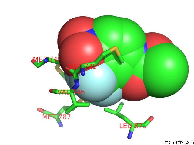
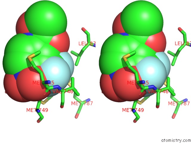
Fluorine binding site 1 out of 3 in 4ogh
Go back to
Fluorine binding site 1 out
of 3 in the Crystal Structure of T877A-Ar-Lbd
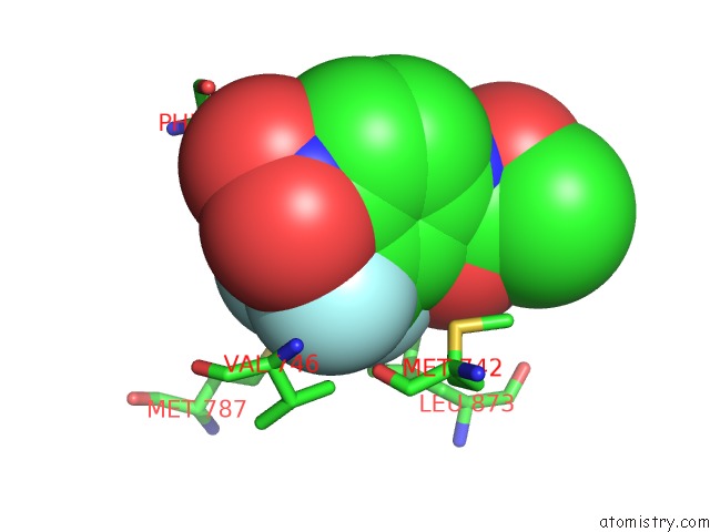
Mono view
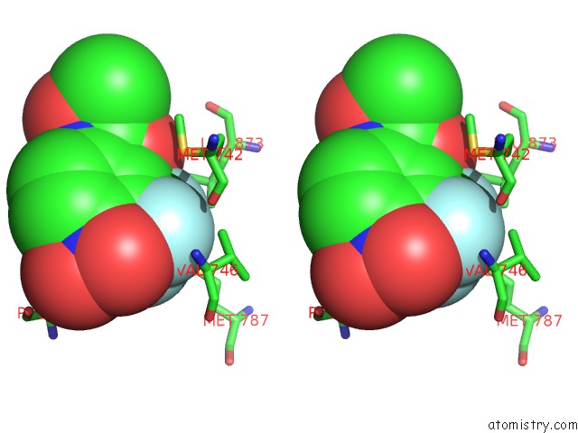
Stereo pair view
Mono view
Stereo pair view
A full contact list of Fluorine with other atoms in the F binding
site number 1 of Crystal Structure of T877A-Ar-Lbd within 5.0Å range:
|
Fluorine binding site 2 out of 3 in 4ogh
Go back to
Fluorine binding site 2 out
of 3 in the Crystal Structure of T877A-Ar-Lbd
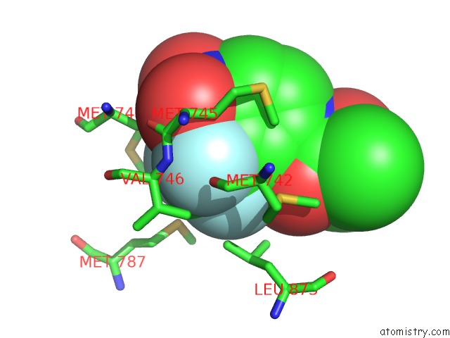
Mono view
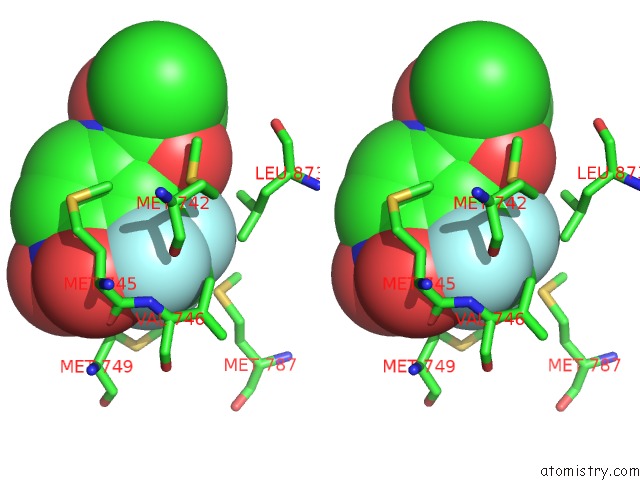
Stereo pair view
Mono view
Stereo pair view
A full contact list of Fluorine with other atoms in the F binding
site number 2 of Crystal Structure of T877A-Ar-Lbd within 5.0Å range:
|
Fluorine binding site 3 out of 3 in 4ogh
Go back to
Fluorine binding site 3 out
of 3 in the Crystal Structure of T877A-Ar-Lbd
Mono view
Stereo pair view
Mono view
Stereo pair view
A full contact list of Fluorine with other atoms in the F binding
site number 3 of Crystal Structure of T877A-Ar-Lbd within 5.0Å range:
|
Reference:
C.L.Hsu,
J.S.Liu,
P.L.Wu,
H.H.Guan,
Y.L.Chen,
A.C.Lin,
H.J.Ting,
S.T.Pang,
S.D.Yeh,
W.L.Ma,
C.J.Chen,
W.G.Wu,
C.Chang.
Identification of A New Androgen Receptor (Ar) Co-Regulator BUD31 and Related Peptides to Suppress Wild-Type and Mutated Ar-Mediated Prostate Cancer Growth Via Peptide Screening and X-Ray Structure Analysis. Mol Oncol 2014.
PubMed: 25091737
DOI: 10.1016/J.MOLONC.2014.06.009
Page generated: Mon Jul 14 23:50:59 2025
PubMed: 25091737
DOI: 10.1016/J.MOLONC.2014.06.009
Last articles
Mg in 2WVMMg in 2WVL
Mg in 2WVJ
Mg in 2WVG
Mg in 2WVA
Mg in 2WTZ
Mg in 2WTY
Mg in 2WTP
Mg in 2WTO
Mg in 2WSS