Fluorine »
PDB 4olm-4pa0 »
4p6g »
Fluorine in PDB 4p6g: Crystal Structure of Human Cathepsin S Bound to A Non-Covalent Inhibitor.
Enzymatic activity of Crystal Structure of Human Cathepsin S Bound to A Non-Covalent Inhibitor.
All present enzymatic activity of Crystal Structure of Human Cathepsin S Bound to A Non-Covalent Inhibitor.:
3.4.22.27;
3.4.22.27;
Protein crystallography data
The structure of Crystal Structure of Human Cathepsin S Bound to A Non-Covalent Inhibitor., PDB code: 4p6g
was solved by
Y.Wang,
P.K.Jadhav,
with X-Ray Crystallography technique. A brief refinement statistics is given in the table below:
| Resolution Low / High (Å) | 65.21 / 1.58 |
| Space group | P 1 |
| Cell size a, b, c (Å), α, β, γ (°) | 67.253, 67.311, 67.778, 100.90, 94.86, 117.99 |
| R / Rfree (%) | 19.8 / 22.2 |
Fluorine Binding Sites:
The binding sites of Fluorine atom in the Crystal Structure of Human Cathepsin S Bound to A Non-Covalent Inhibitor.
(pdb code 4p6g). This binding sites where shown within
5.0 Angstroms radius around Fluorine atom.
In total 4 binding sites of Fluorine where determined in the Crystal Structure of Human Cathepsin S Bound to A Non-Covalent Inhibitor., PDB code: 4p6g:
Jump to Fluorine binding site number: 1; 2; 3; 4;
In total 4 binding sites of Fluorine where determined in the Crystal Structure of Human Cathepsin S Bound to A Non-Covalent Inhibitor., PDB code: 4p6g:
Jump to Fluorine binding site number: 1; 2; 3; 4;
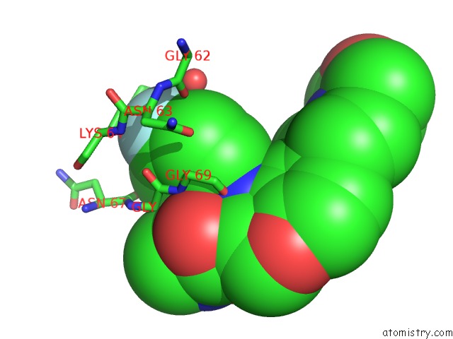
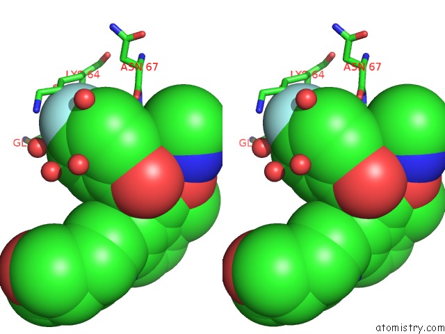
Fluorine binding site 1 out of 4 in 4p6g
Go back to
Fluorine binding site 1 out
of 4 in the Crystal Structure of Human Cathepsin S Bound to A Non-Covalent Inhibitor.
Mono view
Stereo pair view
Mono view
Stereo pair view
A full contact list of Fluorine with other atoms in the F binding
site number 1 of Crystal Structure of Human Cathepsin S Bound to A Non-Covalent Inhibitor. within 5.0Å range:
|
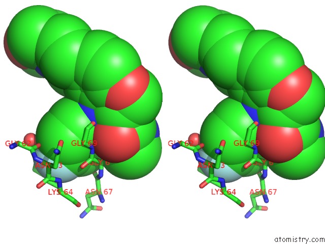
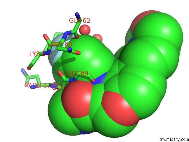
Fluorine binding site 2 out of 4 in 4p6g
Go back to
Fluorine binding site 2 out
of 4 in the Crystal Structure of Human Cathepsin S Bound to A Non-Covalent Inhibitor.

Mono view
Stereo pair view
Mono view
Stereo pair view
A full contact list of Fluorine with other atoms in the F binding
site number 2 of Crystal Structure of Human Cathepsin S Bound to A Non-Covalent Inhibitor. within 5.0Å range:
|
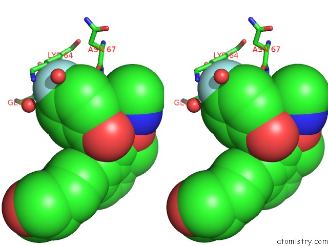
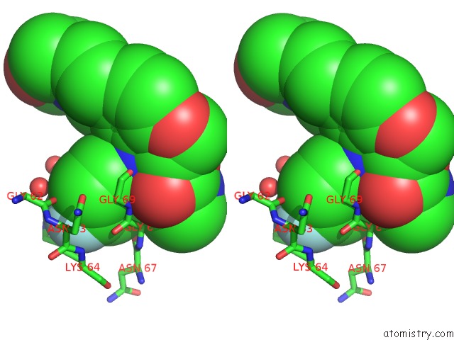
Fluorine binding site 3 out of 4 in 4p6g
Go back to
Fluorine binding site 3 out
of 4 in the Crystal Structure of Human Cathepsin S Bound to A Non-Covalent Inhibitor.
Mono view
Stereo pair view
Mono view
Stereo pair view
A full contact list of Fluorine with other atoms in the F binding
site number 3 of Crystal Structure of Human Cathepsin S Bound to A Non-Covalent Inhibitor. within 5.0Å range:
|
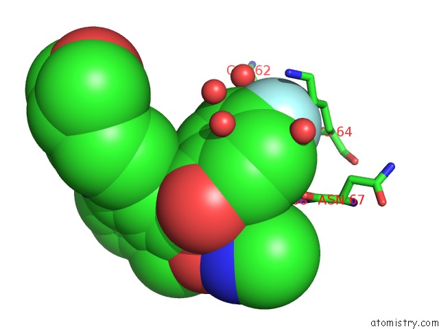
Fluorine binding site 4 out of 4 in 4p6g
Go back to
Fluorine binding site 4 out
of 4 in the Crystal Structure of Human Cathepsin S Bound to A Non-Covalent Inhibitor.
Mono view
Stereo pair view
Mono view
Stereo pair view
A full contact list of Fluorine with other atoms in the F binding
site number 4 of Crystal Structure of Human Cathepsin S Bound to A Non-Covalent Inhibitor. within 5.0Å range:
|
Reference:
P.K.Jadhav,
M.A.Schiffler,
K.Gavardinas,
E.J.Kim,
D.P.Matthews,
M.A.Staszak,
D.S.Coffey,
B.W.Shaw,
K.C.Cassidy,
R.A.Brier,
Y.Zhang,
R.M.Christie,
W.F.Matter,
K.Qing,
J.D.Durbin,
Y.Wang,
G.G.Deng.
Discovery of Cathepsin S Inhibitor LY3000328 For the Treatment of Abdominal Aortic Aneurysm. Acs Med.Chem.Lett. V. 5 1138 2014.
ISSN: ISSN 1948-5875
PubMed: 25313327
DOI: 10.1021/ML500283G
Page generated: Tue Jul 15 00:01:26 2025
ISSN: ISSN 1948-5875
PubMed: 25313327
DOI: 10.1021/ML500283G
Last articles
Ni in 4URHNi in 4V2W
Ni in 4UEQ
Ni in 4V2V
Ni in 4UWX
Ni in 4UQP
Ni in 4URA
Ni in 4UQL
Ni in 4UPV
Ni in 4UPE