Fluorine »
PDB 4qte-4rv6 »
4rm2 »
Fluorine in PDB 4rm2: Crystal Structure of A Benzoate Coenzyme A Ligase with 2-Fluoro Benzoic Acid
Protein crystallography data
The structure of Crystal Structure of A Benzoate Coenzyme A Ligase with 2-Fluoro Benzoic Acid, PDB code: 4rm2
was solved by
S.Strom,
M.Nosrati,
C.Thornburg,
K.D.Walker,
J.H.Geiger,
with X-Ray Crystallography technique. A brief refinement statistics is given in the table below:
| Resolution Low / High (Å) | 41.61 / 1.77 |
| Space group | P 1 21 1 |
| Cell size a, b, c (Å), α, β, γ (°) | 58.885, 95.749, 98.605, 90.00, 110.43, 90.00 |
| R / Rfree (%) | 15.9 / 20.2 |
Fluorine Binding Sites:
The binding sites of Fluorine atom in the Crystal Structure of A Benzoate Coenzyme A Ligase with 2-Fluoro Benzoic Acid
(pdb code 4rm2). This binding sites where shown within
5.0 Angstroms radius around Fluorine atom.
In total 3 binding sites of Fluorine where determined in the Crystal Structure of A Benzoate Coenzyme A Ligase with 2-Fluoro Benzoic Acid, PDB code: 4rm2:
Jump to Fluorine binding site number: 1; 2; 3;
In total 3 binding sites of Fluorine where determined in the Crystal Structure of A Benzoate Coenzyme A Ligase with 2-Fluoro Benzoic Acid, PDB code: 4rm2:
Jump to Fluorine binding site number: 1; 2; 3;
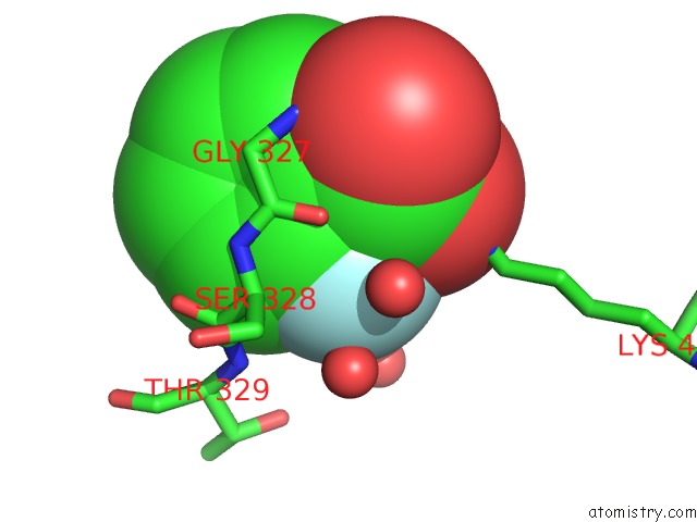
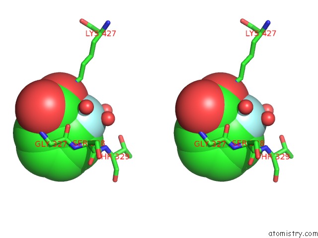
Fluorine binding site 1 out of 3 in 4rm2
Go back to
Fluorine binding site 1 out
of 3 in the Crystal Structure of A Benzoate Coenzyme A Ligase with 2-Fluoro Benzoic Acid
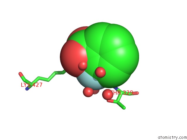
Mono view
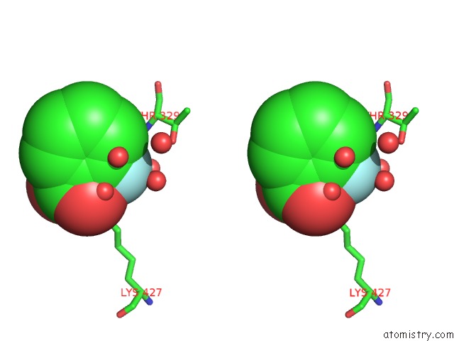
Stereo pair view
Mono view
Stereo pair view
A full contact list of Fluorine with other atoms in the F binding
site number 1 of Crystal Structure of A Benzoate Coenzyme A Ligase with 2-Fluoro Benzoic Acid within 5.0Å range:
|
Fluorine binding site 2 out of 3 in 4rm2
Go back to
Fluorine binding site 2 out
of 3 in the Crystal Structure of A Benzoate Coenzyme A Ligase with 2-Fluoro Benzoic Acid
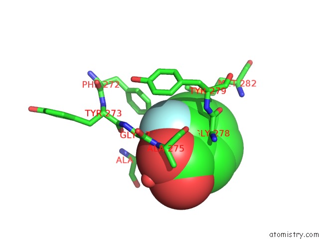
Mono view
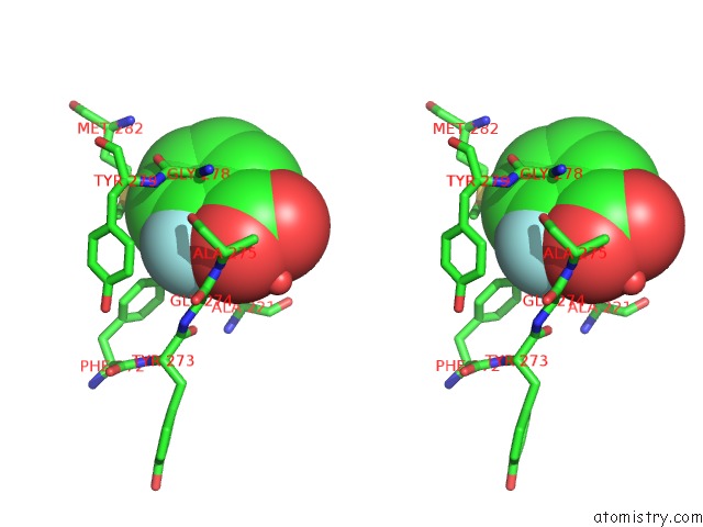
Stereo pair view
Mono view
Stereo pair view
A full contact list of Fluorine with other atoms in the F binding
site number 2 of Crystal Structure of A Benzoate Coenzyme A Ligase with 2-Fluoro Benzoic Acid within 5.0Å range:
|
Fluorine binding site 3 out of 3 in 4rm2
Go back to
Fluorine binding site 3 out
of 3 in the Crystal Structure of A Benzoate Coenzyme A Ligase with 2-Fluoro Benzoic Acid
Mono view
Stereo pair view
Mono view
Stereo pair view
A full contact list of Fluorine with other atoms in the F binding
site number 3 of Crystal Structure of A Benzoate Coenzyme A Ligase with 2-Fluoro Benzoic Acid within 5.0Å range:
|
Reference:
C.K.Thornburg,
S.Wortas-Strom,
M.Nosrati,
J.H.Geiger,
K.D.Walker.
Kinetically and Crystallographically Guided Mutations of A Benzoate Coa Ligase (Bada) Elucidate Mechanism and Expand Substrate Permissivity. Biochemistry V. 54 6230 2015.
ISSN: ISSN 0006-2960
PubMed: 26378464
DOI: 10.1021/ACS.BIOCHEM.5B00899
Page generated: Tue Jul 15 00:39:11 2025
ISSN: ISSN 0006-2960
PubMed: 26378464
DOI: 10.1021/ACS.BIOCHEM.5B00899
Last articles
Na in 5UD3Na in 5UD2
Na in 5UDP
Na in 5UD1
Na in 5UD0
Na in 5UCZ
Na in 5UCS
Na in 5UBU
Na in 5UCQ
Na in 5UCP