Fluorine »
PDB 4rx0-4u7z »
4txn »
Fluorine in PDB 4txn: Crystal Structure of Uridine Phosphorylase From Schistosoma Mansoni in Complex with 5-Fluorouracil
Enzymatic activity of Crystal Structure of Uridine Phosphorylase From Schistosoma Mansoni in Complex with 5-Fluorouracil
All present enzymatic activity of Crystal Structure of Uridine Phosphorylase From Schistosoma Mansoni in Complex with 5-Fluorouracil:
2.4.2.3;
2.4.2.3;
Protein crystallography data
The structure of Crystal Structure of Uridine Phosphorylase From Schistosoma Mansoni in Complex with 5-Fluorouracil, PDB code: 4txn
was solved by
A.Marinho,
J.Torini,
L.Romanello,
A.Cassago,
R.Demarco,
J.Brandao-Neto,
H.M.Pereira,
with X-Ray Crystallography technique. A brief refinement statistics is given in the table below:
| Resolution Low / High (Å) | 29.02 / 2.00 |
| Space group | P 21 21 21 |
| Cell size a, b, c (Å), α, β, γ (°) | 95.643, 107.488, 115.576, 90.00, 90.00, 90.00 |
| R / Rfree (%) | 17.2 / 20.7 |
Fluorine Binding Sites:
The binding sites of Fluorine atom in the Crystal Structure of Uridine Phosphorylase From Schistosoma Mansoni in Complex with 5-Fluorouracil
(pdb code 4txn). This binding sites where shown within
5.0 Angstroms radius around Fluorine atom.
In total 4 binding sites of Fluorine where determined in the Crystal Structure of Uridine Phosphorylase From Schistosoma Mansoni in Complex with 5-Fluorouracil, PDB code: 4txn:
Jump to Fluorine binding site number: 1; 2; 3; 4;
In total 4 binding sites of Fluorine where determined in the Crystal Structure of Uridine Phosphorylase From Schistosoma Mansoni in Complex with 5-Fluorouracil, PDB code: 4txn:
Jump to Fluorine binding site number: 1; 2; 3; 4;
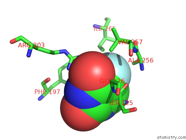
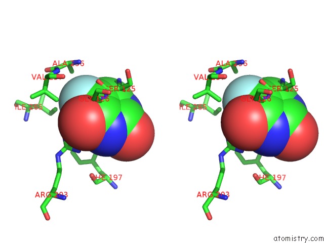
Fluorine binding site 1 out of 4 in 4txn
Go back to
Fluorine binding site 1 out
of 4 in the Crystal Structure of Uridine Phosphorylase From Schistosoma Mansoni in Complex with 5-Fluorouracil
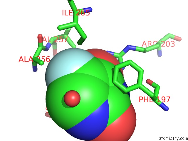
Mono view
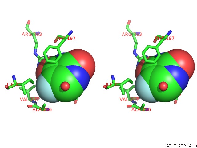
Stereo pair view
Mono view
Stereo pair view
A full contact list of Fluorine with other atoms in the F binding
site number 1 of Crystal Structure of Uridine Phosphorylase From Schistosoma Mansoni in Complex with 5-Fluorouracil within 5.0Å range:
|
Fluorine binding site 2 out of 4 in 4txn
Go back to
Fluorine binding site 2 out
of 4 in the Crystal Structure of Uridine Phosphorylase From Schistosoma Mansoni in Complex with 5-Fluorouracil
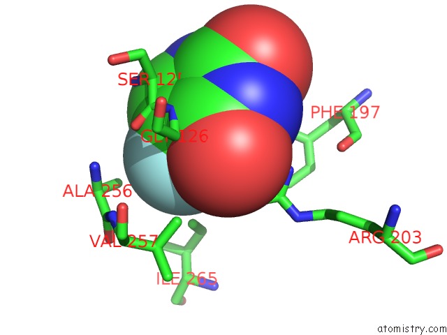
Mono view
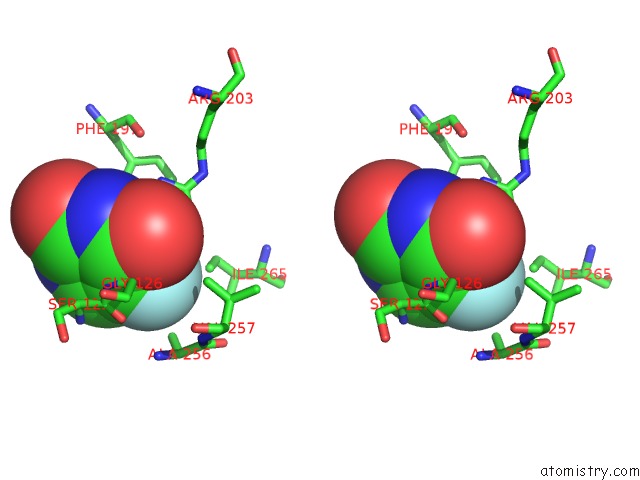
Stereo pair view
Mono view
Stereo pair view
A full contact list of Fluorine with other atoms in the F binding
site number 2 of Crystal Structure of Uridine Phosphorylase From Schistosoma Mansoni in Complex with 5-Fluorouracil within 5.0Å range:
|
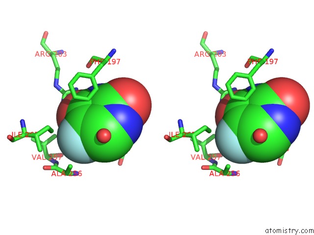
Fluorine binding site 3 out of 4 in 4txn
Go back to
Fluorine binding site 3 out
of 4 in the Crystal Structure of Uridine Phosphorylase From Schistosoma Mansoni in Complex with 5-Fluorouracil
Mono view
Stereo pair view
Mono view
Stereo pair view
A full contact list of Fluorine with other atoms in the F binding
site number 3 of Crystal Structure of Uridine Phosphorylase From Schistosoma Mansoni in Complex with 5-Fluorouracil within 5.0Å range:
|
Fluorine binding site 4 out of 4 in 4txn
Go back to
Fluorine binding site 4 out
of 4 in the Crystal Structure of Uridine Phosphorylase From Schistosoma Mansoni in Complex with 5-Fluorouracil
Mono view
Stereo pair view
Mono view
Stereo pair view
A full contact list of Fluorine with other atoms in the F binding
site number 4 of Crystal Structure of Uridine Phosphorylase From Schistosoma Mansoni in Complex with 5-Fluorouracil within 5.0Å range:
|
Reference:
A.M.Da Silva Neto,
J.R.Torini De Souza,
L.Romanello,
A.Cassago,
V.H.Serrao,
R.Demarco,
J.Brandao-Neto,
R.C.Garratt,
H.D.Pereira.
Analysis of Two Schistosoma Mansoni Uridine Phosphorylases Isoforms Suggests the Emergence of A Protein with A Non-Canonical Function. Biochimie V. 125 12 2016.
ISSN: ISSN 0300-9084
PubMed: 26898674
DOI: 10.1016/J.BIOCHI.2016.02.007
Page generated: Tue Jul 15 00:46:20 2025
ISSN: ISSN 0300-9084
PubMed: 26898674
DOI: 10.1016/J.BIOCHI.2016.02.007
Last articles
Mg in 6O3PMg in 6O2R
Mg in 6O2Q
Mg in 6O36
Mg in 6O2P
Mg in 6O1V
Mg in 6O0J
Mg in 6O1E
Mg in 6O0G
Mg in 6NYY