Fluorine »
PDB 4u80-4uwf »
4uvx »
Fluorine in PDB 4uvx: Crystal Structure of Human Tankyrase 2 in Complex with 3-(4- Chlorophenyl)-5-Fluoro-1,2-Dihydroisoquinolin-1-One
Enzymatic activity of Crystal Structure of Human Tankyrase 2 in Complex with 3-(4- Chlorophenyl)-5-Fluoro-1,2-Dihydroisoquinolin-1-One
All present enzymatic activity of Crystal Structure of Human Tankyrase 2 in Complex with 3-(4- Chlorophenyl)-5-Fluoro-1,2-Dihydroisoquinolin-1-One:
2.4.2.30;
2.4.2.30;
Protein crystallography data
The structure of Crystal Structure of Human Tankyrase 2 in Complex with 3-(4- Chlorophenyl)-5-Fluoro-1,2-Dihydroisoquinolin-1-One, PDB code: 4uvx
was solved by
T.Haikarainen,
M.Narwal,
L.Lehtio,
with X-Ray Crystallography technique. A brief refinement statistics is given in the table below:
| Resolution Low / High (Å) | 29.87 / 1.95 |
| Space group | C 2 2 21 |
| Cell size a, b, c (Å), α, β, γ (°) | 90.580, 98.510, 118.500, 90.00, 90.00, 90.00 |
| R / Rfree (%) | 16.9 / 21.1 |
Other elements in 4uvx:
The structure of Crystal Structure of Human Tankyrase 2 in Complex with 3-(4- Chlorophenyl)-5-Fluoro-1,2-Dihydroisoquinolin-1-One also contains other interesting chemical elements:
| Chlorine | (Cl) | 2 atoms |
| Zinc | (Zn) | 2 atoms |
Fluorine Binding Sites:
The binding sites of Fluorine atom in the Crystal Structure of Human Tankyrase 2 in Complex with 3-(4- Chlorophenyl)-5-Fluoro-1,2-Dihydroisoquinolin-1-One
(pdb code 4uvx). This binding sites where shown within
5.0 Angstroms radius around Fluorine atom.
In total 2 binding sites of Fluorine where determined in the Crystal Structure of Human Tankyrase 2 in Complex with 3-(4- Chlorophenyl)-5-Fluoro-1,2-Dihydroisoquinolin-1-One, PDB code: 4uvx:
Jump to Fluorine binding site number: 1; 2;
In total 2 binding sites of Fluorine where determined in the Crystal Structure of Human Tankyrase 2 in Complex with 3-(4- Chlorophenyl)-5-Fluoro-1,2-Dihydroisoquinolin-1-One, PDB code: 4uvx:
Jump to Fluorine binding site number: 1; 2;
Fluorine binding site 1 out of 2 in 4uvx
Go back to
Fluorine binding site 1 out
of 2 in the Crystal Structure of Human Tankyrase 2 in Complex with 3-(4- Chlorophenyl)-5-Fluoro-1,2-Dihydroisoquinolin-1-One
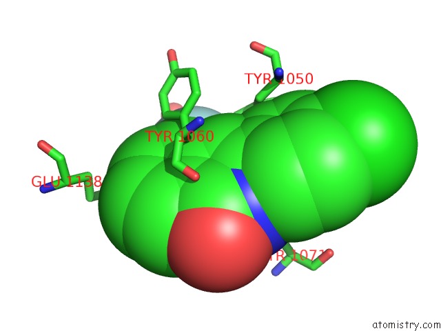
Mono view
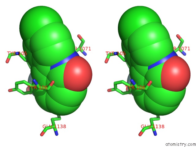
Stereo pair view
Mono view
Stereo pair view
A full contact list of Fluorine with other atoms in the F binding
site number 1 of Crystal Structure of Human Tankyrase 2 in Complex with 3-(4- Chlorophenyl)-5-Fluoro-1,2-Dihydroisoquinolin-1-One within 5.0Å range:
|
Fluorine binding site 2 out of 2 in 4uvx
Go back to
Fluorine binding site 2 out
of 2 in the Crystal Structure of Human Tankyrase 2 in Complex with 3-(4- Chlorophenyl)-5-Fluoro-1,2-Dihydroisoquinolin-1-One
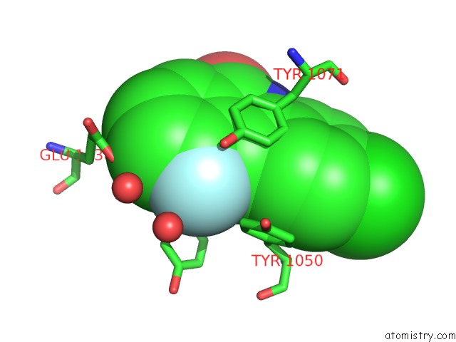
Mono view
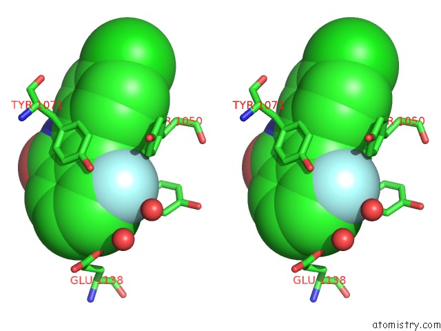
Stereo pair view
Mono view
Stereo pair view
A full contact list of Fluorine with other atoms in the F binding
site number 2 of Crystal Structure of Human Tankyrase 2 in Complex with 3-(4- Chlorophenyl)-5-Fluoro-1,2-Dihydroisoquinolin-1-One within 5.0Å range:
|
Reference:
H.A.Paine,
A.Nathubhai,
E.C.Y.Woon,
P.T.Sunderland,
P.J.Wood,
M.F.Mahon,
M.D.Lloyd,
A.S.Thompson,
T.Haikarainen,
M.Narwal,
L.Lehtio,
M.D.Threadgill.
Exploration of the Nicotinamide-Binding Site of the Tankyrases, Identifying 3-Arylisoquinolin-1-Ones As Potent and Selective Inhibitors in Vitro. Bioorg.Med.Chem. V. 23 5891 2015.
ISSN: ISSN 0968-0896
PubMed: 26189030
DOI: 10.1016/J.BMC.2015.06.061
Page generated: Tue Jul 15 01:06:27 2025
ISSN: ISSN 0968-0896
PubMed: 26189030
DOI: 10.1016/J.BMC.2015.06.061
Last articles
Mg in 2WTZMg in 2WTY
Mg in 2WTP
Mg in 2WTO
Mg in 2WSS
Mg in 2WSB
Mg in 2WPD
Mg in 2WOQ
Mg in 2WOJ
Mg in 2WQS