Fluorine »
PDB 4x7j-4xxs »
4xew »
Fluorine in PDB 4xew: Crystal Structure of 7,8-Diaminopelargonic Acid Synthase (Bioa) From Mycobacterium Tuberculosis, Complexed with A Hts Lead Compound
Enzymatic activity of Crystal Structure of 7,8-Diaminopelargonic Acid Synthase (Bioa) From Mycobacterium Tuberculosis, Complexed with A Hts Lead Compound
All present enzymatic activity of Crystal Structure of 7,8-Diaminopelargonic Acid Synthase (Bioa) From Mycobacterium Tuberculosis, Complexed with A Hts Lead Compound:
2.6.1.62;
2.6.1.62;
Protein crystallography data
The structure of Crystal Structure of 7,8-Diaminopelargonic Acid Synthase (Bioa) From Mycobacterium Tuberculosis, Complexed with A Hts Lead Compound, PDB code: 4xew
was solved by
B.C.Finzel,
R.Dai,
with X-Ray Crystallography technique. A brief refinement statistics is given in the table below:
| Resolution Low / High (Å) | 101.63 / 2.47 |
| Space group | P 21 21 21 |
| Cell size a, b, c (Å), α, β, γ (°) | 62.755, 66.153, 203.261, 90.00, 90.00, 90.00 |
| R / Rfree (%) | 16.5 / 22 |
Fluorine Binding Sites:
The binding sites of Fluorine atom in the Crystal Structure of 7,8-Diaminopelargonic Acid Synthase (Bioa) From Mycobacterium Tuberculosis, Complexed with A Hts Lead Compound
(pdb code 4xew). This binding sites where shown within
5.0 Angstroms radius around Fluorine atom.
In total 2 binding sites of Fluorine where determined in the Crystal Structure of 7,8-Diaminopelargonic Acid Synthase (Bioa) From Mycobacterium Tuberculosis, Complexed with A Hts Lead Compound, PDB code: 4xew:
Jump to Fluorine binding site number: 1; 2;
In total 2 binding sites of Fluorine where determined in the Crystal Structure of 7,8-Diaminopelargonic Acid Synthase (Bioa) From Mycobacterium Tuberculosis, Complexed with A Hts Lead Compound, PDB code: 4xew:
Jump to Fluorine binding site number: 1; 2;
Fluorine binding site 1 out of 2 in 4xew
Go back to
Fluorine binding site 1 out
of 2 in the Crystal Structure of 7,8-Diaminopelargonic Acid Synthase (Bioa) From Mycobacterium Tuberculosis, Complexed with A Hts Lead Compound
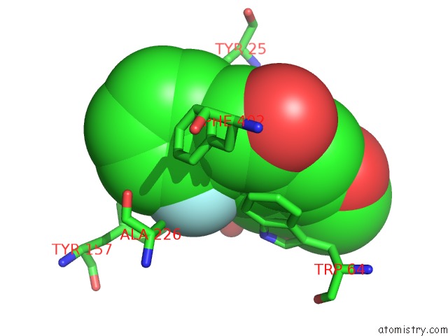
Mono view
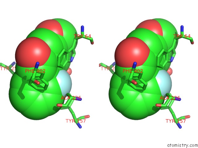
Stereo pair view
Mono view
Stereo pair view
A full contact list of Fluorine with other atoms in the F binding
site number 1 of Crystal Structure of 7,8-Diaminopelargonic Acid Synthase (Bioa) From Mycobacterium Tuberculosis, Complexed with A Hts Lead Compound within 5.0Å range:
|
Fluorine binding site 2 out of 2 in 4xew
Go back to
Fluorine binding site 2 out
of 2 in the Crystal Structure of 7,8-Diaminopelargonic Acid Synthase (Bioa) From Mycobacterium Tuberculosis, Complexed with A Hts Lead Compound
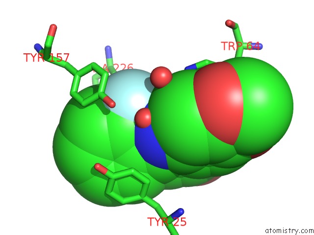
Mono view
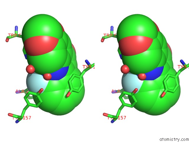
Stereo pair view
Mono view
Stereo pair view
A full contact list of Fluorine with other atoms in the F binding
site number 2 of Crystal Structure of 7,8-Diaminopelargonic Acid Synthase (Bioa) From Mycobacterium Tuberculosis, Complexed with A Hts Lead Compound within 5.0Å range:
|
Reference:
R.Dai,
T.W.Geders,
F.Liu,
S.W.Park,
D.Schnappinger,
C.C.Aldrich,
B.C.Finzel.
Fragment-Based Exploration of Binding Site Flexibility in Mycobacterium Tuberculosis Bioa. J.Med.Chem. V. 58 5208 2015.
ISSN: ISSN 0022-2623
PubMed: 26068403
DOI: 10.1021/ACS.JMEDCHEM.5B00092
Page generated: Tue Jul 15 01:27:53 2025
ISSN: ISSN 0022-2623
PubMed: 26068403
DOI: 10.1021/ACS.JMEDCHEM.5B00092
Last articles
Zn in 9QM9Zn in 9S44
Zn in 9OFE
Zn in 9OFC
Zn in 9OFD
Zn in 9OF1
Zn in 9OFB
Zn in 9N0J
Zn in 9M5X
Zn in 9LGI