Fluorine »
PDB 4xyf-4ymo »
4y2q »
Fluorine in PDB 4y2q: Structure of Soluble Epoxide Hydrolase in Complex with 1-[3- (Trifluoromethyl)Pyridin-2-Yl]Piperazine
Enzymatic activity of Structure of Soluble Epoxide Hydrolase in Complex with 1-[3- (Trifluoromethyl)Pyridin-2-Yl]Piperazine
All present enzymatic activity of Structure of Soluble Epoxide Hydrolase in Complex with 1-[3- (Trifluoromethyl)Pyridin-2-Yl]Piperazine:
3.3.2.10;
3.3.2.10;
Protein crystallography data
The structure of Structure of Soluble Epoxide Hydrolase in Complex with 1-[3- (Trifluoromethyl)Pyridin-2-Yl]Piperazine, PDB code: 4y2q
was solved by
Y.Amano,
T.Yamaguchi,
with X-Ray Crystallography technique. A brief refinement statistics is given in the table below:
| Resolution Low / High (Å) | 76.13 / 2.40 |
| Space group | P 65 2 2 |
| Cell size a, b, c (Å), α, β, γ (°) | 92.544, 92.544, 243.634, 90.00, 90.00, 120.00 |
| R / Rfree (%) | 19.6 / 25.7 |
Other elements in 4y2q:
The structure of Structure of Soluble Epoxide Hydrolase in Complex with 1-[3- (Trifluoromethyl)Pyridin-2-Yl]Piperazine also contains other interesting chemical elements:
| Magnesium | (Mg) | 1 atom |
Fluorine Binding Sites:
The binding sites of Fluorine atom in the Structure of Soluble Epoxide Hydrolase in Complex with 1-[3- (Trifluoromethyl)Pyridin-2-Yl]Piperazine
(pdb code 4y2q). This binding sites where shown within
5.0 Angstroms radius around Fluorine atom.
In total 3 binding sites of Fluorine where determined in the Structure of Soluble Epoxide Hydrolase in Complex with 1-[3- (Trifluoromethyl)Pyridin-2-Yl]Piperazine, PDB code: 4y2q:
Jump to Fluorine binding site number: 1; 2; 3;
In total 3 binding sites of Fluorine where determined in the Structure of Soluble Epoxide Hydrolase in Complex with 1-[3- (Trifluoromethyl)Pyridin-2-Yl]Piperazine, PDB code: 4y2q:
Jump to Fluorine binding site number: 1; 2; 3;
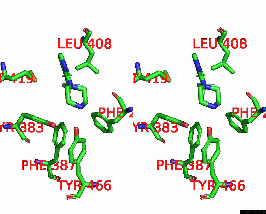
Fluorine binding site 1 out of 3 in 4y2q
Go back to
Fluorine binding site 1 out
of 3 in the Structure of Soluble Epoxide Hydrolase in Complex with 1-[3- (Trifluoromethyl)Pyridin-2-Yl]Piperazine

Mono view

Stereo pair view
Mono view
Stereo pair view
A full contact list of Fluorine with other atoms in the F binding
site number 1 of Structure of Soluble Epoxide Hydrolase in Complex with 1-[3- (Trifluoromethyl)Pyridin-2-Yl]Piperazine within 5.0Å range:
|
Fluorine binding site 2 out of 3 in 4y2q
Go back to
Fluorine binding site 2 out
of 3 in the Structure of Soluble Epoxide Hydrolase in Complex with 1-[3- (Trifluoromethyl)Pyridin-2-Yl]Piperazine

Mono view

Stereo pair view
Mono view
Stereo pair view
A full contact list of Fluorine with other atoms in the F binding
site number 2 of Structure of Soluble Epoxide Hydrolase in Complex with 1-[3- (Trifluoromethyl)Pyridin-2-Yl]Piperazine within 5.0Å range:
|
Fluorine binding site 3 out of 3 in 4y2q
Go back to
Fluorine binding site 3 out
of 3 in the Structure of Soluble Epoxide Hydrolase in Complex with 1-[3- (Trifluoromethyl)Pyridin-2-Yl]Piperazine

Mono view
Stereo pair view
Mono view
Stereo pair view
A full contact list of Fluorine with other atoms in the F binding
site number 3 of Structure of Soluble Epoxide Hydrolase in Complex with 1-[3- (Trifluoromethyl)Pyridin-2-Yl]Piperazine within 5.0Å range:
|
Reference:
Y.Amano,
E.Tanabe,
T.Yamaguchi.
Identification of N-Ethylmethylamine As A Novel Scaffold For Inhibitors of Soluble Epoxide Hydrolase By Crystallographic Fragment Screening Bioorg.Med.Chem. V. 23 2310 2015.
ISSN: ESSN 1464-3391
PubMed: 25862210
DOI: 10.1016/J.BMC.2015.03.083
Page generated: Tue Jul 15 01:37:15 2025
ISSN: ESSN 1464-3391
PubMed: 25862210
DOI: 10.1016/J.BMC.2015.03.083
Last articles
K in 3W1JK in 3WAY
K in 3WAX
K in 3WAW
K in 3WAV
K in 3W8J
K in 3W7A
K in 3W1I
K in 3VUW
K in 3VUX