Fluorine »
PDB 4zga-4zzi »
4zl1 »
Fluorine in PDB 4zl1: Crystal Structure of Human Dihydroorotate Dehydrogenase (Dhodh) with 18X at 1.86 A Resolution
Enzymatic activity of Crystal Structure of Human Dihydroorotate Dehydrogenase (Dhodh) with 18X at 1.86 A Resolution
All present enzymatic activity of Crystal Structure of Human Dihydroorotate Dehydrogenase (Dhodh) with 18X at 1.86 A Resolution:
1.3.5.2;
1.3.5.2;
Protein crystallography data
The structure of Crystal Structure of Human Dihydroorotate Dehydrogenase (Dhodh) with 18X at 1.86 A Resolution, PDB code: 4zl1
was solved by
J.Huang,
D.Wu,
P.Ouyang,
W.Lu,
J.Pu,
with X-Ray Crystallography technique. A brief refinement statistics is given in the table below:
| Resolution Low / High (Å) | 29.04 / 1.86 |
| Space group | P 32 2 1 |
| Cell size a, b, c (Å), α, β, γ (°) | 91.319, 91.319, 122.824, 90.00, 90.00, 120.00 |
| R / Rfree (%) | 16 / 17.9 |
Fluorine Binding Sites:
The binding sites of Fluorine atom in the Crystal Structure of Human Dihydroorotate Dehydrogenase (Dhodh) with 18X at 1.86 A Resolution
(pdb code 4zl1). This binding sites where shown within
5.0 Angstroms radius around Fluorine atom.
In total only one binding site of Fluorine was determined in the Crystal Structure of Human Dihydroorotate Dehydrogenase (Dhodh) with 18X at 1.86 A Resolution, PDB code: 4zl1:
In total only one binding site of Fluorine was determined in the Crystal Structure of Human Dihydroorotate Dehydrogenase (Dhodh) with 18X at 1.86 A Resolution, PDB code: 4zl1:
Fluorine binding site 1 out of 1 in 4zl1
Go back to
Fluorine binding site 1 out
of 1 in the Crystal Structure of Human Dihydroorotate Dehydrogenase (Dhodh) with 18X at 1.86 A Resolution
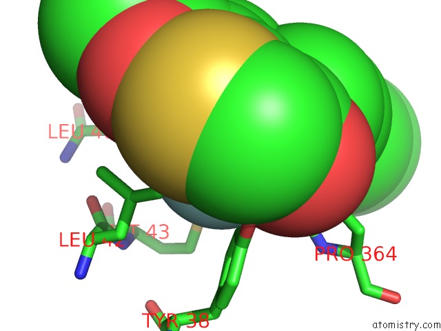
Mono view
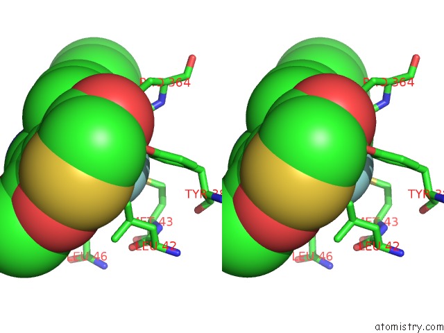
Stereo pair view
Mono view
Stereo pair view
A full contact list of Fluorine with other atoms in the F binding
site number 1 of Crystal Structure of Human Dihydroorotate Dehydrogenase (Dhodh) with 18X at 1.86 A Resolution within 5.0Å range:
|
Reference:
J.Huang,
D.Wu,
P.Ouyang,
W.Lu,
J.Pu.
Crystal Structure of Human Dihydroorotate Dehydrogenase (Dhodh) with 18XYW at 1.86 A Resolution To Be Published.
Page generated: Tue Jul 15 01:58:39 2025
Last articles
Na in 4UUANa in 4UU8
Na in 4UU5
Na in 4UU7
Na in 4UTZ
Na in 4UTX
Na in 4USZ
Na in 4USW
Na in 4UTV
Na in 4UTR