Fluorine »
PDB 4zga-4zzi »
4zth »
Fluorine in PDB 4zth: Structure of Human P38AMAPK-Arylpyridazinylpyridine Fragment Complex Used in Inhibitor Discovery
Enzymatic activity of Structure of Human P38AMAPK-Arylpyridazinylpyridine Fragment Complex Used in Inhibitor Discovery
All present enzymatic activity of Structure of Human P38AMAPK-Arylpyridazinylpyridine Fragment Complex Used in Inhibitor Discovery:
2.7.11.24;
2.7.11.24;
Protein crystallography data
The structure of Structure of Human P38AMAPK-Arylpyridazinylpyridine Fragment Complex Used in Inhibitor Discovery, PDB code: 4zth
was solved by
V.L.Grum-Tokars,
S.M.Roy,
D.M.Watterson,
with X-Ray Crystallography technique. A brief refinement statistics is given in the table below:
| Resolution Low / High (Å) | 30.00 / 2.15 |
| Space group | P 21 21 21 |
| Cell size a, b, c (Å), α, β, γ (°) | 65.767, 74.501, 77.669, 90.00, 90.00, 90.00 |
| R / Rfree (%) | 18.1 / 24 |
Other elements in 4zth:
The structure of Structure of Human P38AMAPK-Arylpyridazinylpyridine Fragment Complex Used in Inhibitor Discovery also contains other interesting chemical elements:
| Chlorine | (Cl) | 1 atom |
Fluorine Binding Sites:
The binding sites of Fluorine atom in the Structure of Human P38AMAPK-Arylpyridazinylpyridine Fragment Complex Used in Inhibitor Discovery
(pdb code 4zth). This binding sites where shown within
5.0 Angstroms radius around Fluorine atom.
In total only one binding site of Fluorine was determined in the Structure of Human P38AMAPK-Arylpyridazinylpyridine Fragment Complex Used in Inhibitor Discovery, PDB code: 4zth:
In total only one binding site of Fluorine was determined in the Structure of Human P38AMAPK-Arylpyridazinylpyridine Fragment Complex Used in Inhibitor Discovery, PDB code: 4zth:
Fluorine binding site 1 out of 1 in 4zth
Go back to
Fluorine binding site 1 out
of 1 in the Structure of Human P38AMAPK-Arylpyridazinylpyridine Fragment Complex Used in Inhibitor Discovery
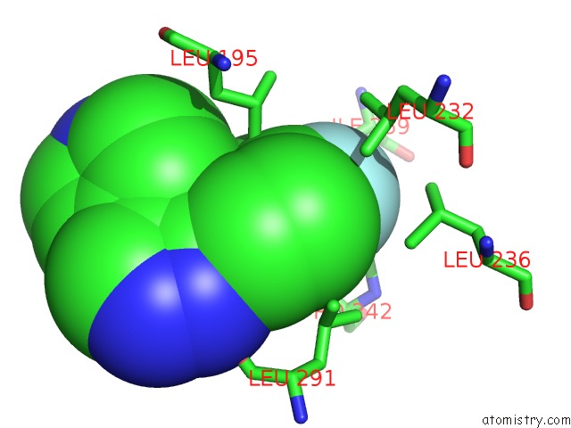
Mono view
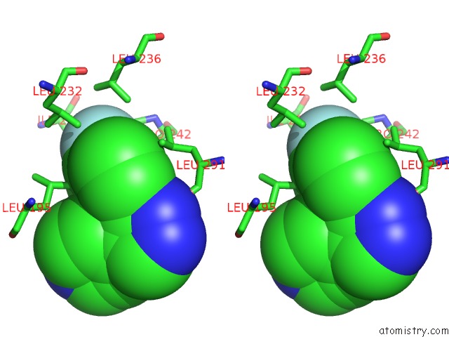
Stereo pair view
Mono view
Stereo pair view
A full contact list of Fluorine with other atoms in the F binding
site number 1 of Structure of Human P38AMAPK-Arylpyridazinylpyridine Fragment Complex Used in Inhibitor Discovery within 5.0Å range:
|
Reference:
V.L.Grum-Tokars,
S.M.Roy,
D.M.Watterson.
Structure of Human P38AMAPK-Arylpyridazinylpyridine Scaffold Complex Used in Inhibitor Discovery To Be Published.
Page generated: Tue Jul 15 02:02:51 2025
Last articles
Na in 4TMENa in 4TMV
Na in 4TLK
Na in 4TM6
Na in 4TLH
Na in 4TLS
Na in 4TKX
Na in 4TL5
Na in 4RYF
Na in 4RU4